
|
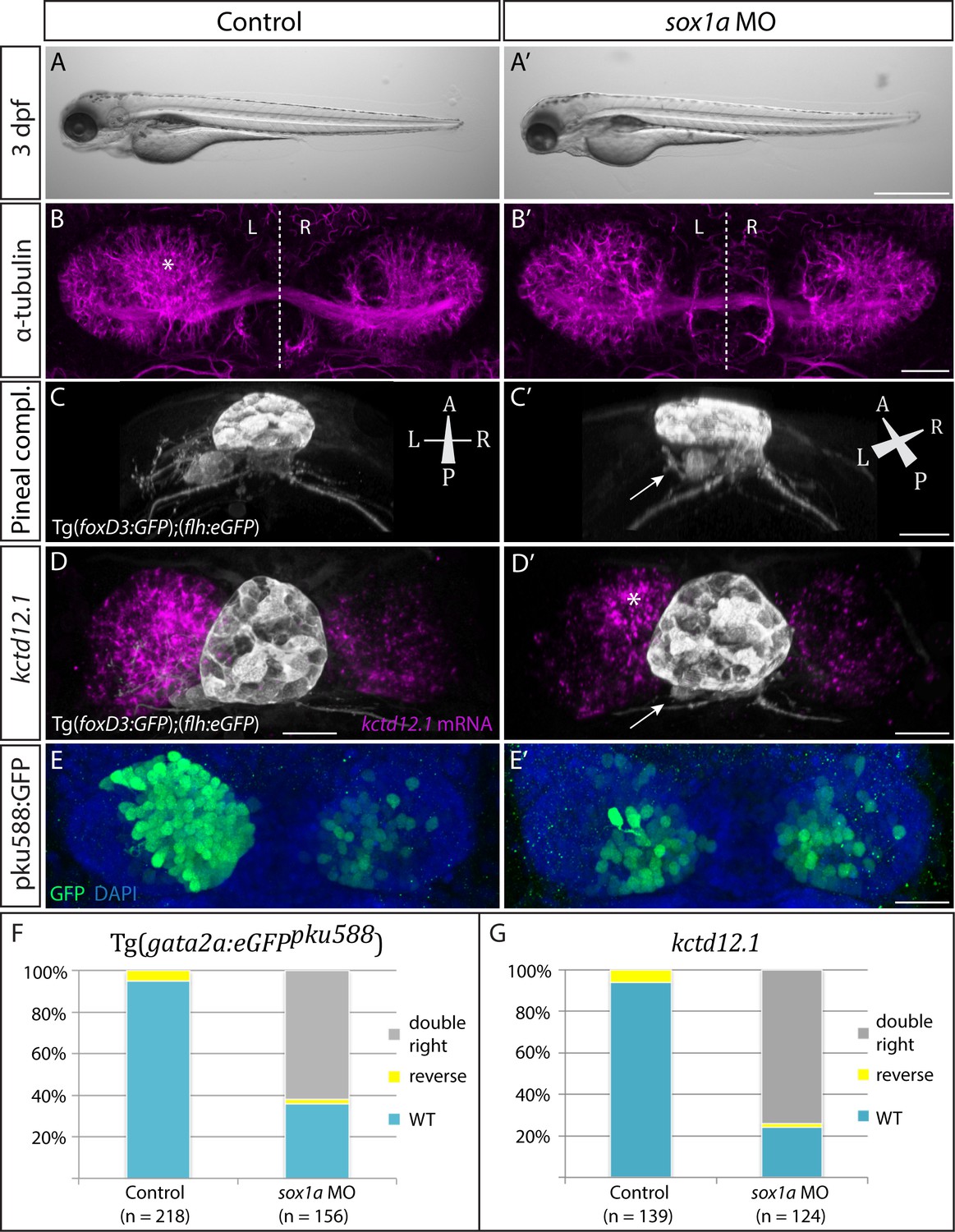
Fig. 3-S1
sox1a morphants show a double-right habenular phenotype comparable to sox1a mutants.
(A–A’) sox1a morpholino (MO)-injected embryo showing normal overall morphology at 3 dpf. Scale bar 500 µm. (B–B’) Dense left dHb neuropil domain (asterisk) is reduced in sox1a morphants at 4 dpf (acetylated α-tubulin immunostaining). Scale bar 25 µm. (C–C’) MO injections into Tg(foxD3:GFP);(flh:eGFP) transgenic fish reveal that the parapineal migrates to its normal position next to the pineal stalk by 4 dpf in the sox1a morphant, but has shortened axonal projections (arrow in C’). Scale bar 25 µm. (D–D’) Left habenula kctd12.1 mRNA expression at 4 dpf is decreased in the sox1a morphant despite normal parapineal migration (arrow), with only a small residual asymmetric domain (asterisk). Scale bar 25 µm. (E–E’) Numbers of GFP-positive neurons in Tg(gata2a:eGFPpku588) line labelling the left-dominant lateral domains of the dHb (located medially in larval zebrafish) is reduced in the 4 dpf sox1a morphant. Scale bar 25 µm. (F–G) Percentages of larvae with double-right, wild-type and reversed habenular phenotypes in controls and upon sox1a MO injections. Scoring is based on Tg(gata2a:eGFPpku588) GFP expression (E–E’, F) and kctd12.1 mRNA expression (D–D’, G) at 4 dpf.