
|
Fig. 5-S1
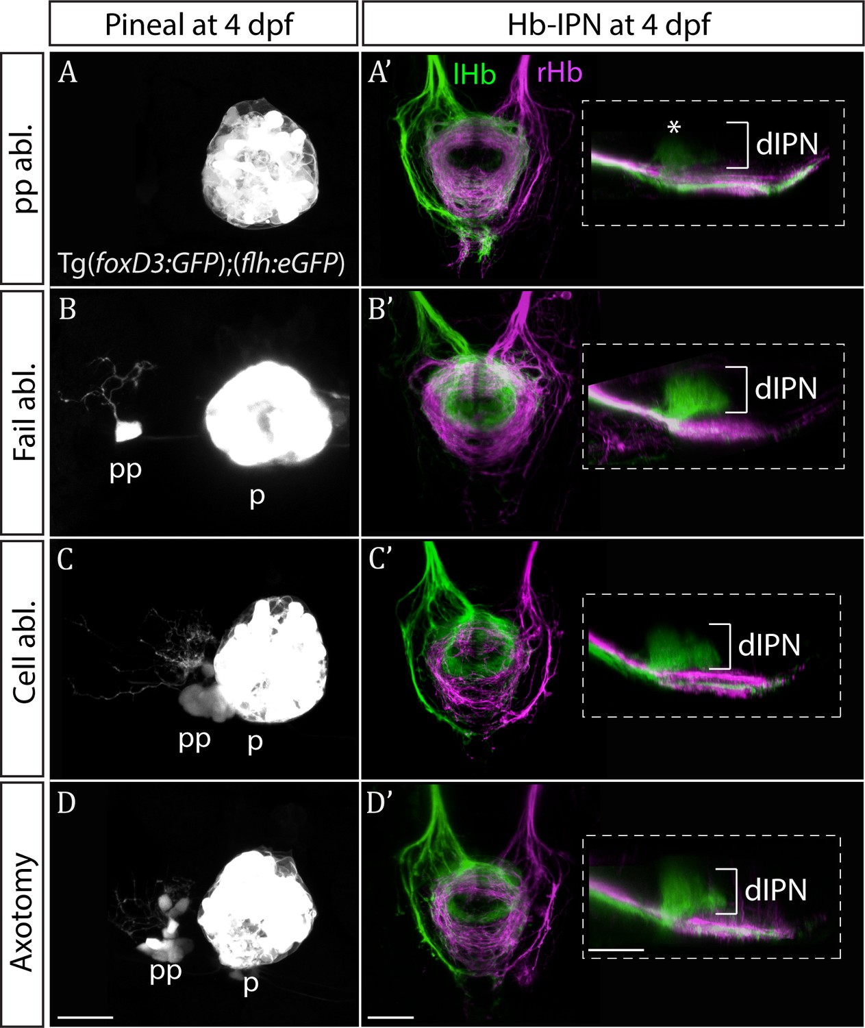
Only ablation of all parapineal cells leads to loss of habenular asymmetries.
(A–D) Live-imaging of the pineal complex at 4 dpf showing expression of Tg(foxD3:GFP)zf104 and Tg(flh:eGFP)U711 transgenes in the pineal (p) and the parapineal (pp). Parapineal ablations were carried out at 50 hpf, parapineal axotomies repeatedly at 50, 60 and 72 hpf. Fail ablation (n = 7) demarks one or two parapineal cells left unablated (B), cell ablation (n = 4) is ablation of 2–3 parapineal cells (C) and axotomies (n = 5) were carried out by severing the parapineal axon bundle approximately 10 µm from the cell bodies, which often also disrupted the compact structure of the parapineal (D). (A’–D’) Dorsal and lateral views of the interpeduncular nucleus labelled by anterograde tracing of the axons from the left dorsal habenula (lHb, green) and right dorsal habenula (rHb, magenta) at 4 dpf. Note that innervation of dorsal interpeduncular nucleus (dIPN) from the left habenula is lost only upon ablation of all parapineal cells. The asterisk indicates a small tuft of neurites in the dorsal anterior IPN that are present upon complete parapineal ablation (Bianco et al., 2008) and can also be detected in sox1a mutants (Figure 3E’). All scale bars 25 µm.