
|
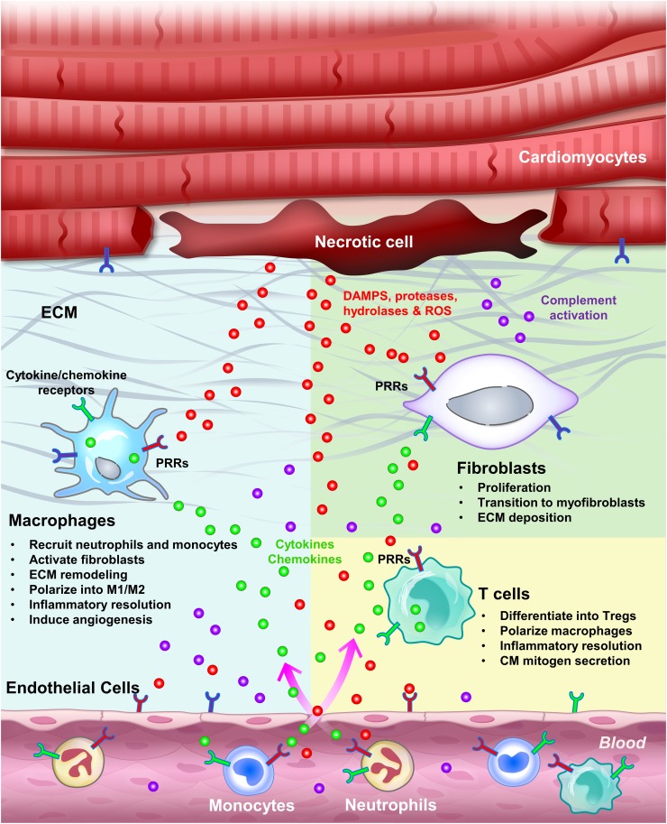
Fig. 2
Inflammation induced by cardiac injury. Sterile inflammation can be triggered by various components released by necrotic cells, including DAMPs, proteases, hydrolases and mitochondrial ROS. DAMPs directly activate PRRs on surveillant cells, including tissue macrophages, circulating monocytes and neutrophils, as well as on resident cells, including endothelial cells, fibroblasts and CMs. Proteases, hydrolases and ROS activate the complement system as well as inflammasomes, and degrade the ECM, altogether further propagating the inflammatory response. Activated tissue resident macrophages secrete cytokines to attract monocytes and neutrophils, activate endothelial cells to promote cell adhesion and permeability, and remodel the ECM. Infiltrating monocytes and neutrophils clear cell debris by phagocytosis and help terminate the initial insult. After wound clearance, myofibroblasts secrete ECM to help prevent the injured heart from rupturing. Differentiated Tregs tune down the inflammation by secreting anti-inflammatory cytokines, in parallel with M1–M2 macrophage polarization and programmed neutrophil apoptosis. Inflammation initiation, propagation and resolution can occur in both regenerative and non-regenerative models. However, in the regenerative models, these processes seem to facilitate CM dedifferentiation and proliferation and scar resolution by mechanisms yet to be determined