
Fig. 3
|
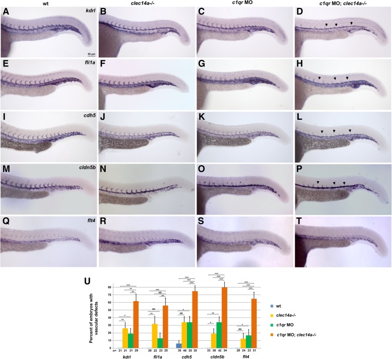
Fig. 3
Analysis of vascular marker expression by in situ hybridization at 24 hpf in clec14a−/− embryos injected with 10 ng of c1qr MO. a-d kdrl, e-h fli1a, i-l cdh5, m-p arterial marker cldn5b, q-t venous marker flt4expression. Note the strong inhibition of angiogenic sprouting in the double clec14a−/−; c1qr MO embryos (arrowheads). Only weak or partial inhibition of sprouting is observed in clec14a−/− or c1qrMO embryos. u Percentage of embryos with defects in ISV sprouting is greatly increased among c1qrMO; clec14a−/− embryos compared to c1qr MO or clec14a−/− embryos. n refers to the number of embryos analyzed for each marker. *p < 0.05; ** p < 0.01; *** p < 0.001; NS, no significance, Fisher’s exact test. Data were combined from two independent experiments. Error bars show standard error