
|
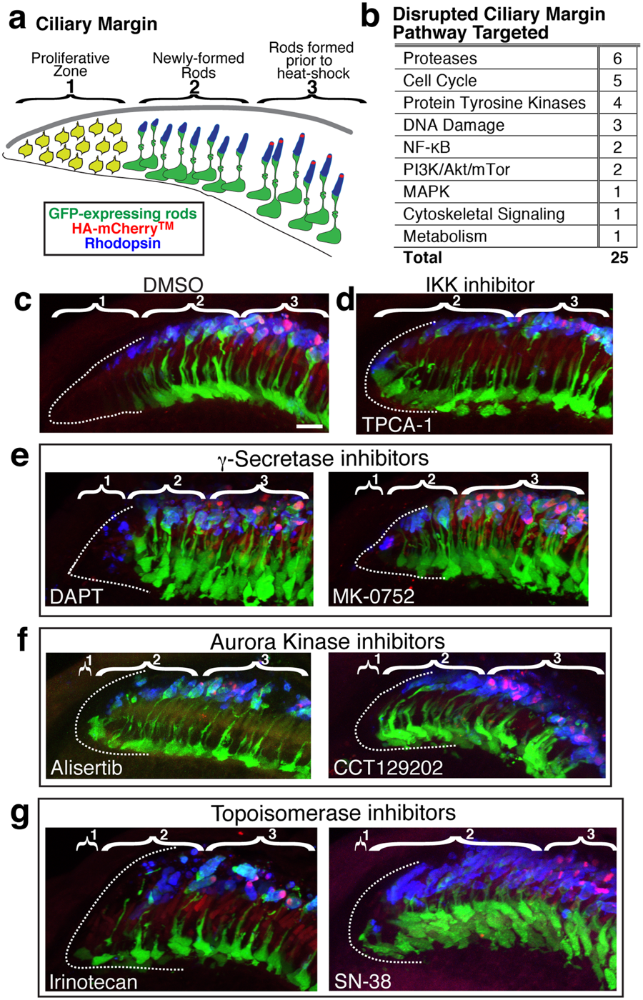
Fig. 6
Notch signaling, cell cycle, and DNA damage pathways are among those targeted by compounds that disrupt the peripheral rim of the retina. (a) The peripheral rim of the retina consists of (1) a proliferative zone, the CMZ, where stem and precursor cells reside, (2) a region of newly-formed retinal neurons and glia including GFP-positive rods, and (3) rods that have the mCherryTM stripe because they developed prior to heat shock. (b) Pathways targeted by compounds that disrupt the peripheral rim of the retina. (c) The peripheral rim from a control DMSO-treated larva segmented into region (1) with dotted white outline, (2) with GFP (green) and Rhodopsin (blue)-positive but mCherryTM stripe-negative rods, and (3) with GFP, Rhodopsin, and mCherryTM (red)-positive rods. (d) The proliferative zone (1) is nearly absent following treatment with IKK inhibitor TPCA-1. (e) The proliferative zone (1) is reduced in size and newly formed rods (2) are packed tightly with γ-Secretase inhibitor (DAPT and MK-0752) treatment. (f) The proliferative zone (1) is dramatically reduced with Aurora Kinase inhibitor (Alisertib and CCT129202) treatment. (g) The proliferative zone is dramatically reduced with topoisomerase inhibitor (Irinotecan and SN-38) treatment. Scale bar is 10 μm.