
|
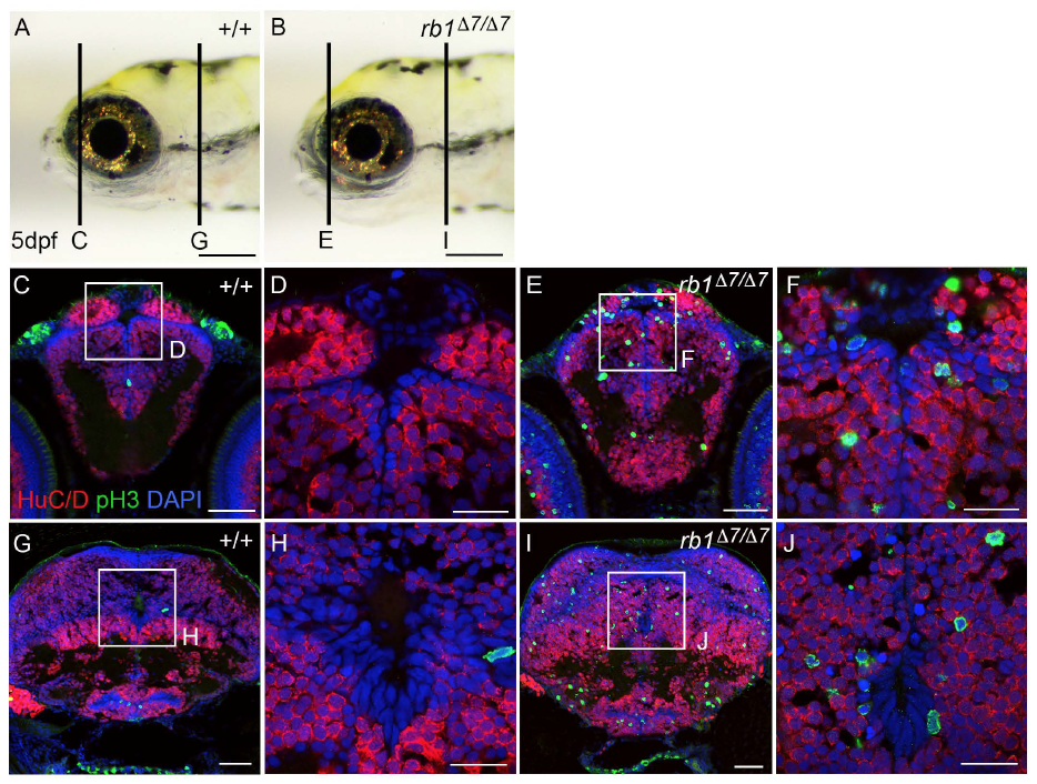
Fig. S7
Ectopic M-phase phosphohistone-H3 positive cells in the telencephalon/forebrain and hindbrain of rb1D7/D7 mutant 5 dpf larva. A wildtype +/+ and B rb1D7/D7 mutant 5 dpf gross morphology. C-J Cryosections were labeled with antibodies to the neuronal marker HuC/D (red) and mitotic M-phase marker phosphohistone-H3 (green). Sections through the telencephalon (C-F) and hindbrain (G-J) show that in wild type mitotic cells are absent or rare, but in the rb1D7/D7 mutant many ectopic phosphohistone-H3 positive cells are scattered throughout the region containing HuC/D-positive cell bodies of neurons.