
|
Fig. 7
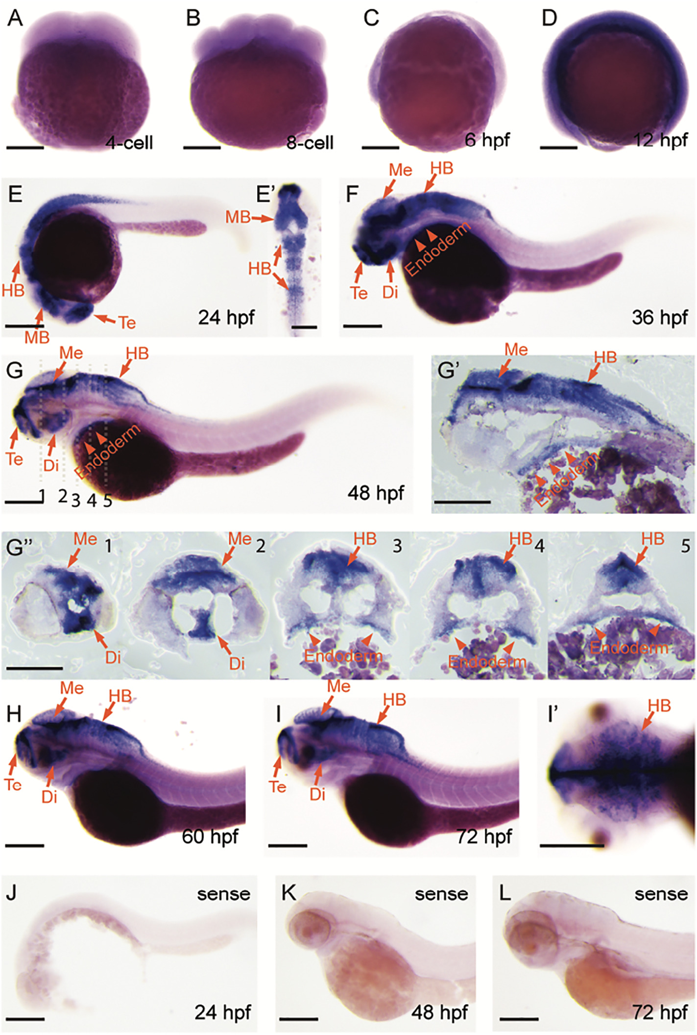
Expression pattern of gene fgfbp3 in zebrafish embryo. Whole-mount in situ hybridization of fgfbp3 mRNA at 4-cell stage (A), 8-cell stage (B), 6hpf (C), 12hpf (D), 24hpf (E and E′), 36hpf (F), 48hpf (G, G′, and G″), 60hpf (H), and 72hpf (I and I′). (A–I′) Antisense probe. (J–L) Sense probe. (A, B, C, D, E, F, G, H, I, and J–L) Lateral view. (E′) Flat mount of 24hpf embryo, and dorsal view. (G′) Longitudinal section of 48hpf embryos in panel (G), White dashed lines in panel (G) denote plane of cross section in panels (G″1–5). Abbreviations: Di, diencephalon; HB, hindbrain; MB, midbrain; Me, mesencephalon; Te, telencephalon. Scale bar: 200 μm.
Reprinted from Gene, 659, Li, Y., Sun, S., Ding, Z., Yang, C., Zhang, G., Jiang, Q., Zou, Y., Temporal and spatial expression of fgfbp genes in zebrafish, 128-136, Copyright (2018) with permission from Elsevier. Full text @ Gene