
Image
|
Figure Caption
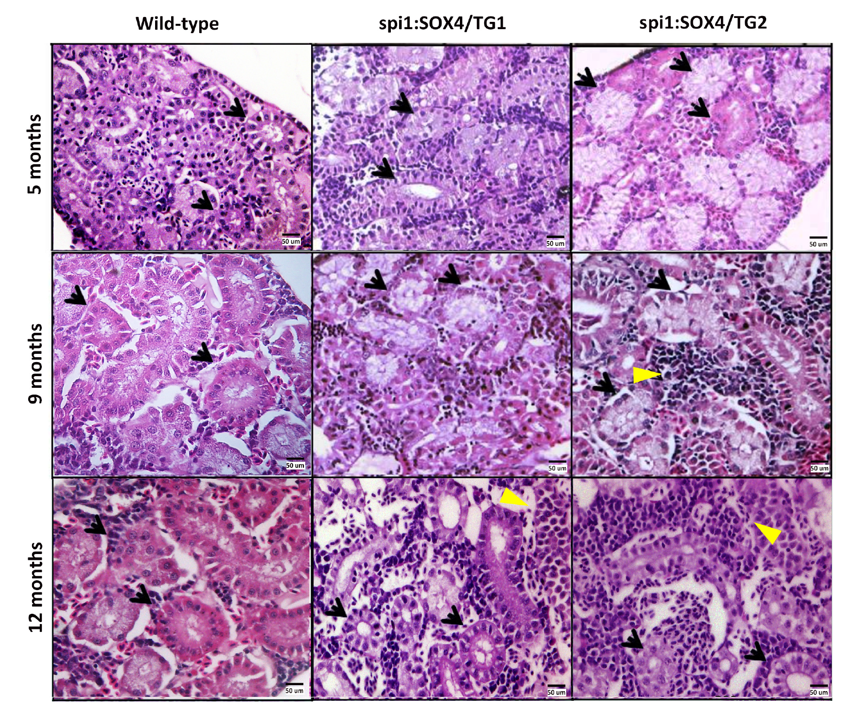
Fig. S1
Histological analysis of the kidney of Tg(spi1:SOX4-EGFP) zebrafish. H&E stains of the kidney are from 5-, 9- and 12-month-old Tg(spi1:SOX4-EGFP) and wild-type fish. The kidney from spi1:SOX4-EGFP fish showed infiltration by myeloid cells at 9 and 12 -months. Arrow: renal tubule. Yellow triangle: infiltration cells. Magnification: 400x.
Figure Data
Acknowledgments
This image is the copyrighted work of the attributed author or publisher, and
ZFIN has permission only to display this image to its users.
Additional permissions should be obtained from the applicable author or publisher of the image.
Full text @ Blood Cancer J