
|
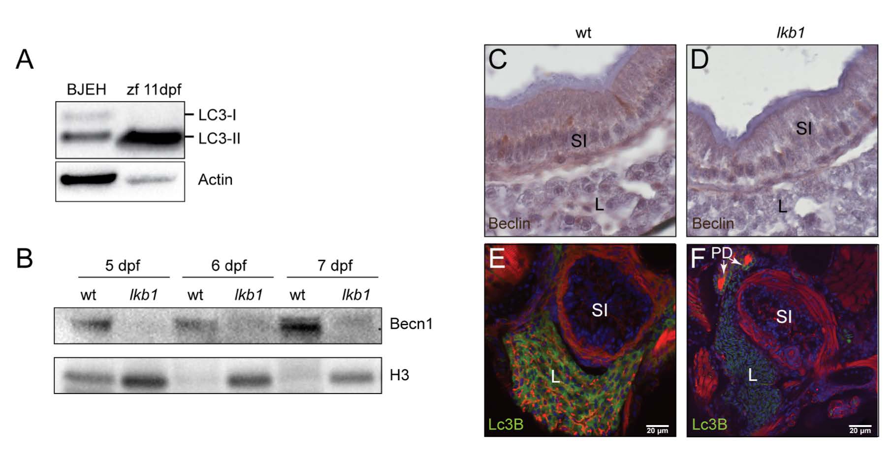
Fig. S2
Autophagy markers Lc3B and Beclin are lower in lkb1 mutants. (A) The LC3B antibody recognizes the cleaved Lc3-II in zebrafish. Western blot analysis using antibodies against LC3B and beta-actin (loading control) on total protein lysates from human BJEH cells that were serum-starved overnight and wt zebrafish larvae at 11 dpf. The LC3B antibody recognizes the uncleaved and cleaved forms of LC3B in the human sample, but predominantly the cleaved Lc3-II in the zebrafish sample. Uncropped images of the blots are presented in Supplementary Fig. S13. (B) Western blot analysis of Beclin (Becn1) and Histone H3 (loading control) in total protein lysates of wt and lkb1 trunks between 5-7 dpf. Larvae were treated with chloroquine (2.5 μM) for 14 h prior to processing. Becn1 levels are lower in the lkb1 mutants at all time-points. (C-D) Immunohistochemical analysis of transverse paraffin sections (5 μm) of intestine of 7 dpf wt and lkb1 larvae shows very low levels of Becn1 expression in the lkb1 intestine. Magnification: 100X. (E-F) Transverse vibratome sections (150 μm) of liver of 7 dpf wt and lkb1 mutants stained with anti-LC3B antibody (green), rhodamine-phalloidin to detect F-actin (red), and DAPI to detect nuclei (blue). Lc3B staining in the lkb1 liver is greatly reduced. PD: pronephric ducts; L: liver; SI: intestine.