
|
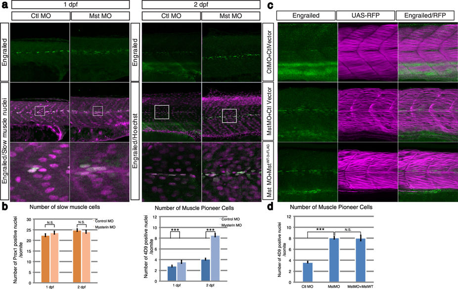
Fig. 5
Suppression of mysterin-α significantly increases the number of MPCs.
(a) Increased number of MPCs upon suppression of mysterin-α. MPCs in the trunk region are stained with an anti-engrailed antibody (green). The nuclei of slow muscle cells at 1 dpf and all cells at 2 dpf are stained with an anti-prox1 antibody (magenta, left panels) and Hoechst (magenta, right panels), respectively. The morphant has a significantly increased number of MPCs at 2 dpf but not at 1 dpf. (b) Quantification of slow muscle cells and MPCs. The left panel shows the mean number of nuclei of slow muscle cells (prox1-positive) within 20, 34, 49, and 33 somites of control animals at 1 dpf, mysterin-α morphants at 1 dpf, control animals at 2 dpf, and mysterin-α morphants at 2 dpf, respectively. The right panel shows the mean number of slow muscle nuclei (engrailed-positive) within 43, 52, 20, and 34 somites of control animals at 1 dpf, mysterin-α morphants at 1 dpf, control animals at 2 dpf, and mysterin-α morphants at 2 dpf, respectively. Error bars represent the standard deviation. N.S.: not significant. ***P < 0.001. (c) Effect of fast muscle-specific expression of human mysterin on the number of MPCs. Human mysterin tagged with a 3×FLAG epitope (WT-3×FLAG) is expressed in embryonic fast muscle using the Tol2 and GAL4/UAS systems. RFP expression is driven by fast muscle-specific GAL4FF and is thus an indicator of GAL4 activity and fast muscle morphology (See Methods). The number of engrailed-positive nuclei (green) in morphants is unchanged by fast muscle-specific expression of human mysterin. (d) Quantification of MPCs shown in (c). The panel shows the mean number of nuclei of MPCs (engrailed-positive) within 64, 29, and 25 somites of control animals, mysterin-α morphants, and human mysterin-expressing morphants, respectively, at 2 dpf. Error bars represent the standard deviation. N.S.: not significant. ***P < 0.001.