
|
Fig. 3
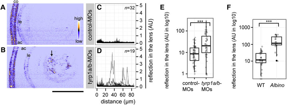
Absence of mature melanosomes sensitizes the lens fibre to cataract formation.
(A,B) Confocal reflection imaging on the lenses at 4 dpf of living embryos injected with either control-MOs (A) or tyrp1a/b-MOs (B). The anterior chamber (ac) is oriented to the left. co: cornea. le: lens epithelium. Scale bar: 50 μm. The arrow in panel B points out abnormal lens reflection observed after tyrp1a/b-knockdown. (C,D) Lenticular reflection profiles as a function of distance from the anterior edge of the lens capsule (lc) toward the posterior end of the lens for control-MO injected embryos (C) and tyrp1a/b-MO injected embryos (D). The intensity of reflection is shown in an arbitrary unit (AU). The number of examined individuals for each group is shown in the upper right corner. Profiles from the individual embryos were overlaid. (E,F) Quantification of abnormal lens reflections. Welch two sample t-test and Mann-Whitney U test showed significant differences between control-MOs (n = 51 embryos) and tyrp1a/b-MO injections (n = 40 embryos; (E) ***p = 8.74 × 10⁻⁴, t = 3.5896), and between wild type embryos (WT, n = 29 embryos) and albino (slc45a2) homozygous embryos (n = 19 embryos; (F) ***p = 1.52 × 10⁻⁷, U = 26), respectively.