
|
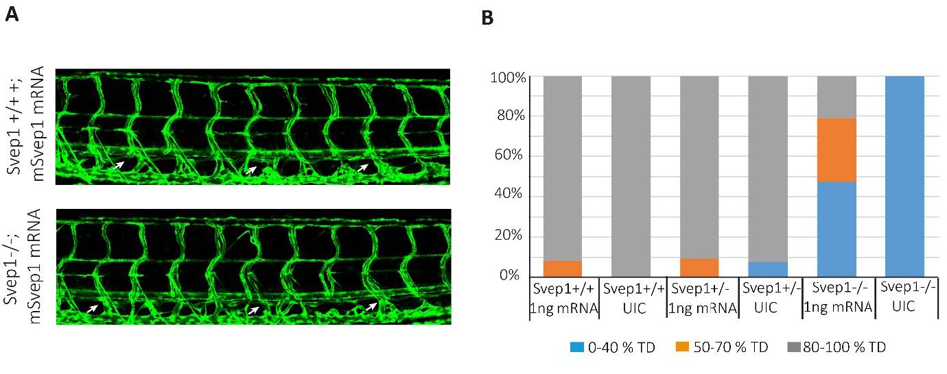
Fig. S2
Rescue of svep1 mutants with murine Svep1 mRNA: A: Lateral view of polydom/svep1 sibling (top) and mutant (bottom) embryos at 5dpf. Embryos had been injected at the 1-2 cell stage with 1ng of murine Svep1 mRNA generated by in vitro transcription. At 5dpf the extent of the TD (arrows) across ten body segments was scored (arrows), and embryos were subsequently genotyped. Note the complete thoracic duct structure in the mutant embryo. B: Quantification of a separate experiment, where control embryos were left un-injected, while experimental embryos were injected with 1ng of murine Svep1 mRNA. Embryos were scored as described above, and subsequently genotyped. All un-injected mutant embryos developed thoracic duct fragments in maximally 40% of their trunk segments, while in injected embryos 30% showed a thoracic duct in 50-70% of their trunk segments, and 20% of injected mutant embryos even showed complete rescue.