
|
Fig. S1
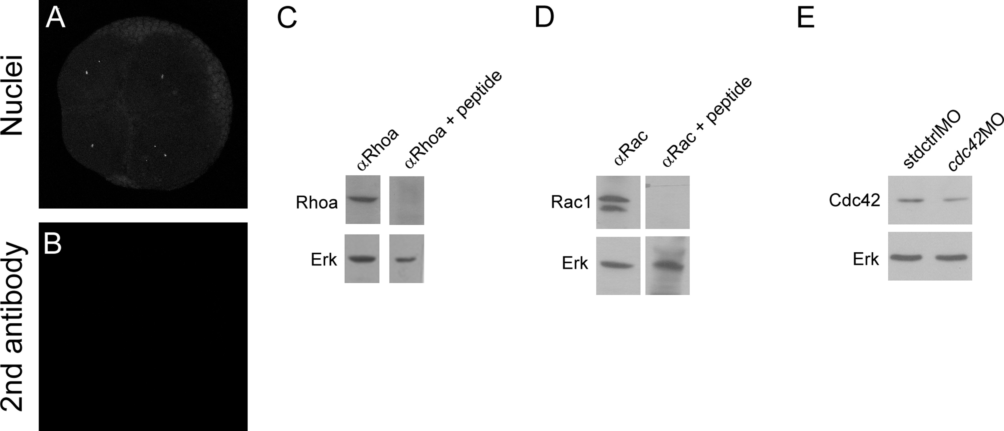
Rhoa, Cdc42 and Rac1 antibodies recognize their respective proteins. Embryos at 4 to 8- cell transition were co-stained with DAPI to show the nuclei (A) and does not show any positive signal after inmunodetection with only anti-rabbit secondary antibody (B). Whole protein samples were obtained from 24 hpf embryos. A single band of the expected size is observed in the western blot against RhoA (C top left panel). Incubation of the primary RhoA antibody with RhoA specific peptide competes and the band in the western blot is not longer observed (C top right panel). Membranes were stripped and blotted against Erk as loading control (C bottom left and right panels). A doublet is observed in western blot against Rac1 (D top left panel) that was competed with specific peptide against Rac1 (D top right panel), membranes were stripped and blotted against Erk as loading control (D bottom left and right panel). Cdc42 specificity was demonstrated by using specific morpholino against cdc42 (cdc42MO). The amount of Cdc42 is less in cdc42 morfants compared to embryos injected with standard morpholino (stdMO). Membranes were stripped and blotted against Erk as a loading control. Western blots experiments were repeated at least in 6 times in independent experiments and antibodies validation were repeated 3 times.
Reprinted from Developmental Biology, 421(1), Miranda-Rodríguez, J.R., Salas-Vidal, E., Lomelí, H., Zurita, M., Schnabel, D., RhoA/ROCK pathway activity is essential for the correct localization of the germ plasm mRNAs in zebrafish embryos, 27-42, Copyright (2017) with permission from Elsevier. Full text @ Dev. Biol.