
|
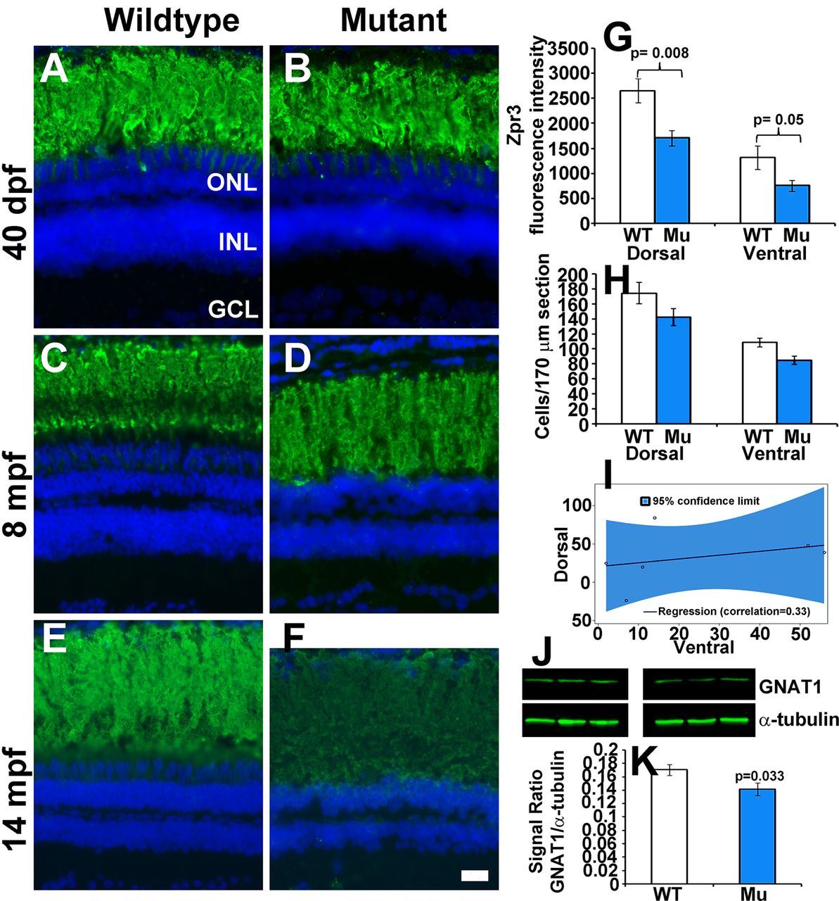
Fig. 6
Rod photoreceptors degenerate in older EYS-deficient zebrafish. (A-F) Retinal sections were stained with Zpr3 and counter stained with DAPI. (A,B) Zpr3 staining of wild-type and EYS-deficient zebrafish at 40 dpf. (C,D) Zpr3 staining of wild-type and EYS-deficient zebrafish at 8 mpf. (E,F) Zpr3 staining of wild-type and EYS-deficient zebrafish at 14 mpf. (G) Quantification of Zpr3 immunofluorescence intensity of wild-type and EYS-deficient zebrafish at 14 mpf. (H,I) Rod quantification in wild-type and EYS-deficient zebrafish at 14 mpf. Rod cells in dorsal and ventral retina near the optic nerve heads were counted on H&E stained sections from sex- and body weight-matched animals. (J,K) GNAT1 western blot and quantification at 14 mpf. Western blot was carried out with GNAT1 and α-tubulin antibodies. Note that no significant differences were observed between wild-type and EYS-deficient zebrafish at 40 dpf and 8 mpf. However, Zpr3 immunoreactivity, rod cell numbers and GNAT1 protein were reduced in EYS-deficient zebrafish at 14 mpf. Scale bar for A-F: 20 µm. Error bars in G,H,K indicate s.e.m.