
|
Fig. 5
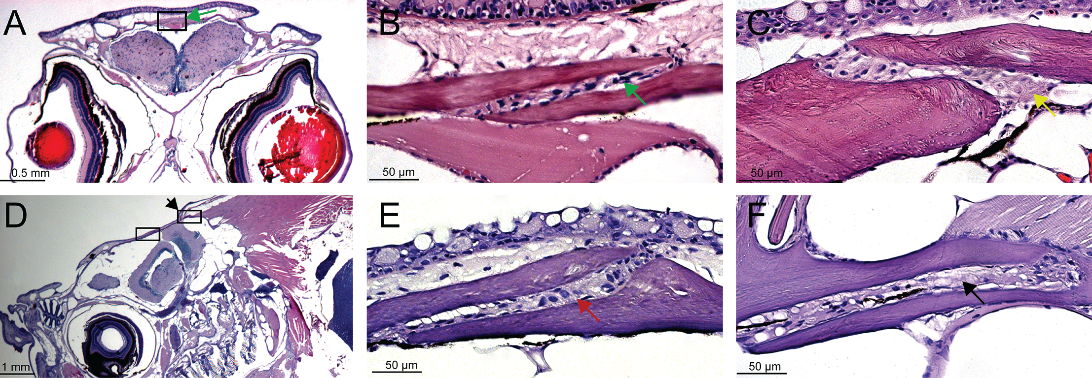
Histological examination of cranial sutures in adult zebrafish.
H&E stained transverse sections (A-C) and sagittal sections (D-E) of skulls. (A) The interfrontal suture (boxed) and presented in (B) higher magnification; green arrows indicate the interfrontal suture between overlapping frontal bones. (C) The sagittal suture (yellow arrow) observed between two parietal bones. (D) The sagittal plane of sectioning revealed the coronal (left box) and lambdoid sutures (right box, black arrow). (E) The posterior frontal bone overlapping the anterior portion of the parietal bone, with the coronal suture formed between them (red arrow). (F) The lambdoid suture (black arrow) separates the parietal (upper plate) and the supraoccipital bone (lower plate).