
|
Fig. 5
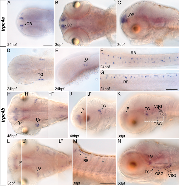
Expression patterns of trpc4 paralogs during development. A-C: Expression of trpc4a was exclusively found in the olfactory bulb. Dorsal (A,B) and lateral (C) views of representative embryonic and larval stages are shown. D-G: Images showing trpc4b staining in embryonic head (D,E) and tail (F,G) region 24 hr postfertilization (hpf) were taken dorsally (D,F) and laterally (E,G). H,J: Whole-mount zebrafish 48 hpf, shown in dorsal (H) and lateral (J) views. Prime figures show the same embryo in different focal planes with H′′ focusing on hindbrain expression. K-M: Lateral (K,M) and dorsal (L) views on trpc4b expression in 3 days postfertilization (dpf) whole-mount larvae. Dotted box in K has a more lateral focal plane compared with core image. L-L′′: different focal planes of the same larva with L′ and L′′ focusing on trigeminal and hindbrain expression, respectively. N: Expression of trpc4b spreads to all cranial sensory ganglia around 5 dpf. Anterior is always left. FSG, facial sensory ganglia; GSG, glossopharyngeal sensory ganglia; OB, olfactory bulb; P, pallium; RB, Rohon-Beard neurons; TG, trigeminal ganglia; VSG, vagal sensory ganglia. Scale bar = 100 µm in A (applies to all images if not otherwise indicated), F,G,M.