
|
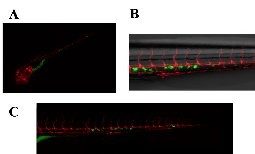
Fig. 3
Images of carboxyfluorescein succinimidyl ester (CFSE)-labeled, patient-derived breast cancer bone metastasis (primary cells) xenografted in Tg(kdrl:mcherry) ZF embryos. (A) Whole-body image of the zebrafish embryo at 5 dpi (25× magnification). Primary cells disseminated predominantly in the caudal hematopoietic tissues (CHT) of the embryo; (B) Details of (A), showing the interactions of primary cells with the zebrafish vessels in the CHT. Cells extravasated in the CHT of the ZF embryo and engrafted in close proximity of the vessels. Fluorescent images merged with brightfield image, 63× magnification; (C) Combined picture of the CHT of embryo in (A,B). Individual images were taken at 63× magnification.