
|
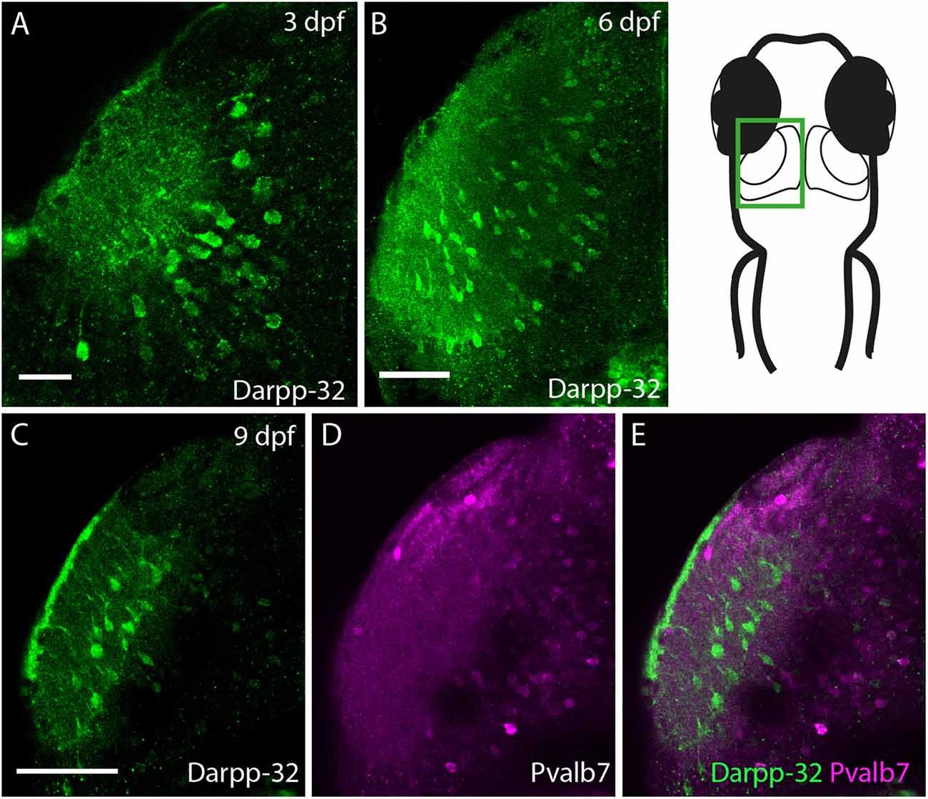
Fig. 10
Darpp-32 expression in the larval optic tectum. Single optical sections of whole mount immunofluorescence stainings. The images are showing one hemisphere of the optic tectum as indicated in the schematic on the right side. (A) Darpp-32 expression in the optic tectum can be first seen at 3 dpf. Darpp-32 is expressed in the neuropil and the cell bodies of projection neurons. (B) Darpp-32 expression in optic tectum at 6 dpf. (C-E) Note that during the course of development the expression of Pvalb7 (D) and Darpp-32 (C) does not overlap in the optic tectum (E). In (C) intense Darpp-32 expression is visible in the superficial most layer, presumably the SM, while Pvalb 7 expression is detectable more closer to the midline, presumably in neurons of the stratum periventriculare (SPV). Darpp-32 positive cell bodies located are more lateral compared to Pvalb 7 positive ones. Scale bar in (A) 20 µm; in (B-E) 50 µm.