
|
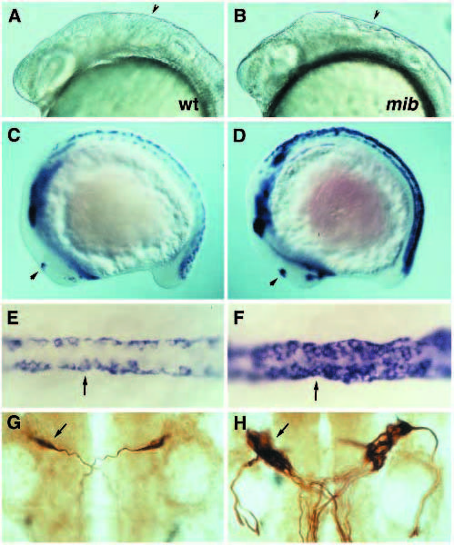
Fig. 9
Phenotypic analysis of mind bomb (mib) mutants. (A,C,E,G) Wild type. (B,D,F,H) mind bomb (mib)m178 mutants. (A,B) Brain in wild-type (A) and mutant (B) embryos at 25-somite stage. Note the irregularities in the hindbrain (arrowhead). (C,D) Expression of islet1 in wild-type (C) and mutant (D) embryos at 13-somite stage. Note the dramatic increase of islet1-expressing cells in all regions where primary neurons are formed, including the spinal cord, epiphysis (arrow) and trigeminal ganglion. (E,F) Dorsal view of prospective Rohon-Beard cells (arrow) in dorsal spinal cord of embryos shown in C and D. (G,H) Mauthner neurons (arrow) in wild type (G) and mutant (H) embryos at 28 hpf, highlighted by 3A10 antibody. Note the abnormal projection of the most laterally located supernumerary Mauthner cell in this mutant embryo. All the other Mauthner cells project towards the midline.