
|
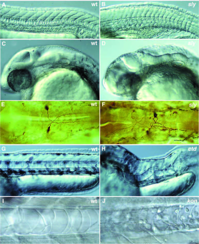
Fig. 8
Photographs of a variety of selected mutants with late notochord defects. (A-D) Lateral views of 1- day old wild-type and mutant zebrafish embryos. (A,C) 1-day old wild-type embryo. (B,D) 1-day old slyti263a. (A) In wild type, the notochord cells are vacuolated and the notochord has a ‘stack-of-pennies’ appearance. (B) In embryos with strong alleles of sly, the notochord is thinner and poorly vacuolated. In a lateral view, the brain of sly embryos looks very disorganized when compared to wild type (C,D). (E,F) Antibody staining of the Mauthner neurons in 1-day old embryos using the monoclonal antibody 3A10. (E) In wild type, the axons of the Mauthner neurons cross the midline and grow towards the posterior. (F) In sly, the axons of one or both Mauthner neurons frequently fail to cross the midline and grow posteriorly along the ipsilateral side. (G,H) Lateral views of 2-day old wild-type and mutant embryos. (G) Lateral view of wild type with a normal, straight notochord. (H) Lateral view of ctd, in which the notochord extends in a random fashion throughout the embryo. (I) Notochord of a 40-hour old wildtype embryo with an intact notochord sheath. (J) Notochord of a 40-hour old kontc230 embryo. The vacuolated notochord cells are surrounded by undifferentiated round cells that failed to become notochord sheath cells.