
|
Fig. 3
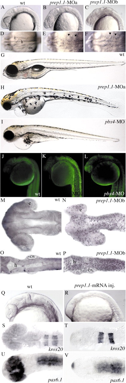
The phenotype of prep1.1 morphants is characterized by apoptosis. (A-I) 24-hpf and 5-dpf embryos. (B,C,E,F) Embryos injected with either prep1.1-MOa or prep1.1-MOb have similar phenotypes, characterized by alterations in brain morphology and widespread cell death, which is clearly visible as a higher opacity that is particularly evident at the level of the hindbrain (arrowheads in E,F). (H) prep1.1 morphants are characterized by small head and eyes, lack of jaws, abnormal distribution of melanocytes and delayed reabsorption of the yolk sac. (I) pbx4 morphants are characterized by small head and eyes and reduced jaws. Both prep1.1 and pbx4 morphants display a swollen pericardium and lack an inflated swim bladder. (A-C,G-I) are lateral, and (D-F) are dorsal views, with anterior to the left. (J-L) Acridine orange staining reveals widespread cell death in prep1.1 morphants, but not in wild-type embryos and pbx4 morphants. Embryos are in lateral views. (M-P) TUNEL labelling reveals intense DNA fragmentation in the brain of prep1.1 morphants at the 22-somite stage, which is more pronounced in the hindbrain. Embryos are in dorsal views with anterior to the left. (Q-V) Injection of prep1.1 mRNA posteriorizes the zebrafish embryo. Visual inspection (Q,R), and in situ hybridization with krox20 (S,T) and pax6.1 (U,V) antisense probes reveals that embryos injected with prep1.1 mRNA (R,T,V) have a reduced forebrain compared to uninjected embryos (Q,S,U). e, eye; h, hindbrain; I, isthmus; ov, otic vesicle.