
|
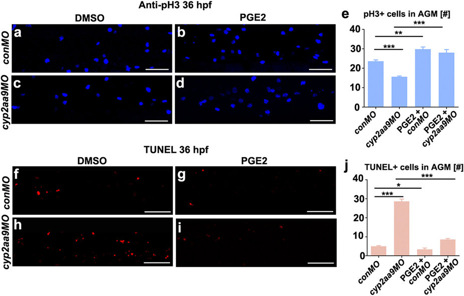
Fig. 4
Defective proliferation and apoptosis caused by cyp2aa9MO were rescued by PGE2.
(a-d) cyp2aa9MO led to reduced number of mitotic active cells at 36 hpf in the AGM as shown by pH3 antibody staining, which could be rescued by PGE2. (e) Quantification of pH3-positive cells in the AGM (n = 6, mean ± SD). (f-i) cyp2aa9MO led to increases in the number of apoptotic cells at 36 hpf in the AGM as shown by TUNEL assay, which could be rescued by PGE2. (j) Quantification of the number of apoptotic cells in the AGM (n = 6, mean ± SD). *p < 0.05, **p < 0.01, ***p < 0.001, Student’s t-test. All the views are exactly the AGM area shown in lateral, anterior left, dorsal top. Scale bar, 50 µm.