
|
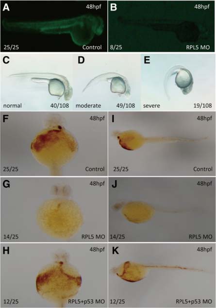
Fig. 1
Hemoglobin staining of embryos injected with RPL5 MO and the effectiveness of RPL5 MO. a-b Embryos co-injected with 25 ng RPL5:egfp DNA and 0.25 ng control MO produced green fluorescent protein (a), and the expression of the green fluorescent fusion protein was inhibited by co-injection with 0.25 ng RPL5 MO (b). c-e The different phenotype and its ratio of embryos injected with 0.25 ng RPL5 MO f-k The O-staining results revealed a drastic reduction in the number of hemoglobin-stained blood cells when RPL5 was knocked down (f and i show the control; g and j show the RPL5 knockdown), and this was partially rescued by coinjection with P53 MO (h and k). a, b, c, d, e,i, j and k show the lateral view; f, g, and h show the ventral view