
Fig. 5
|
Fig. 5
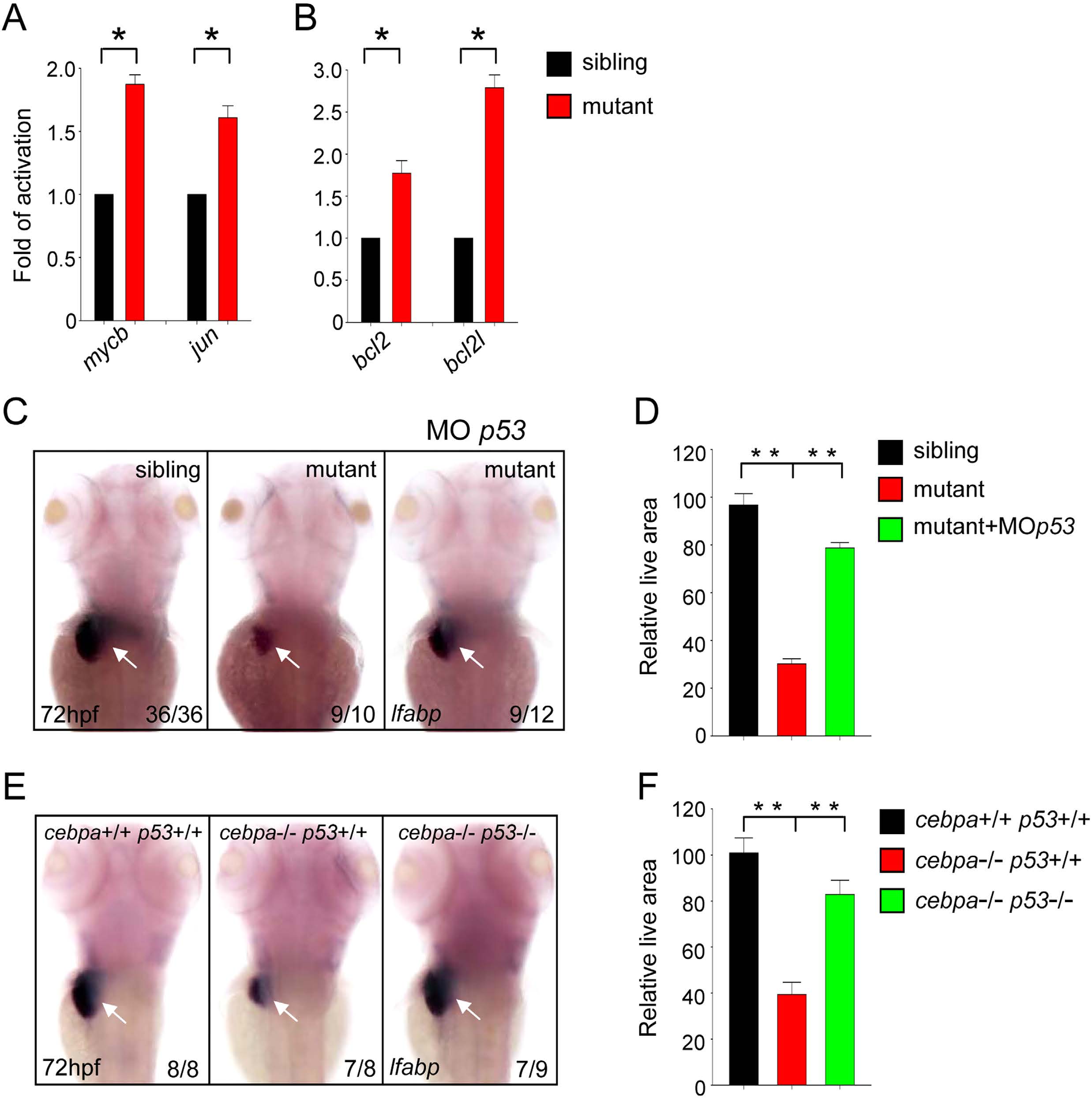
The p53 signaling pathway is activated in cebpa-deficient developing liver.
(A–B) Quantitative PCR analysis of the expression of cell proliferation and apoptosis-related genes in 72 hpf embryos. Data shown are the mean ± SD, n e 3, *P < 0.05 by student’s t-test. (C,E) WISH assay of lfabp at 72 hpf. Loss of p53 could rescue the hepatic defect in cebpa mutant embryos. Dorsal views with anterior to the top. (D,F) The relative liver area measured in (C,E) respectively. The result shown is fold difference compared with the level (set to 100) detected in control embryos (mean ± SD, n e 3, **P < 0.005 by student’s t-test).