
|
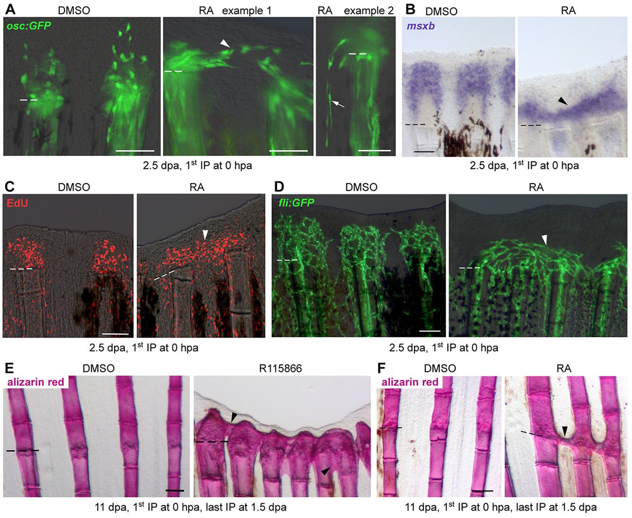
Fig. 2
Preosteoblast alignment and maintenance of the ray-interray organization requires RA-degrading epidermal niches. (A) IHC for GFP in osc:gfp fish reveals ectopic GFP+ preosteoblasts in interrays in RA-treated fish at 2.5dpa. Arrowhead indicates preosteoblast in interray tissue, arrow indicates proximally migrating preosteoblast in interray tissue. (B,C) WISH for msxb (B) and EdU labeling (C) at 2.5dpa demonstrate expansion of blastema cells into interrays in RA-treated fish. Arrowheads indicate ectopic blastema cells. (D) fli:gfp fish show misconnected blood vessels (arrowhead) in RA-treated fish at 2.5dpa. (E,F) RA and R115866 treatment during blastema formation result in formation of ectopic bone at the wound site and in R115866-treated fish in an irreversible regeneration block. Arrowheads indicate ectopic bone, dashed lines indicate amputation plane. Scale bars: 100µm.