
|
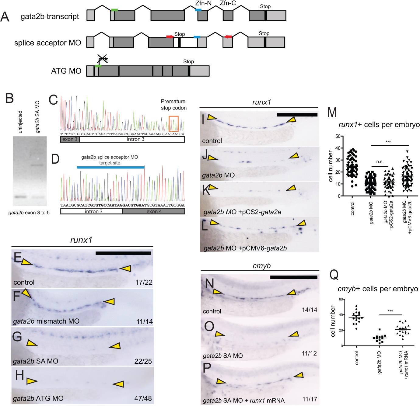
Fig. S3 gata2b morpholino validation. (A) Schematic representation of gata2b morpholino targets. Light grey boxes indicate untranslated regions (UTRs). Zfn-N and Zfn-C indicate the location of the N- and C-terminal zinc finger domains. The gata2b splice acceptor morpholino, represented by a blue bar, targets the intron 3 / exon 4 boundary upstream of the zinc finger DNA binding domains. Red arrows indicate the positioning of primers designed to validate the splice acceptor morpholino. A gata2b ATG morpholino, represented in (A) by a green bar, is designed to block translation of gata2b transcript. (B) RT-PCR of gata2b in embryos injected with gata2b splice acceptor morpholino. Primers amplify transcript from exon 3 to 5. (C-D) Representative sequences of the splice product produced by the gata2b splice acceptor MO. (E-H) In situ hybridization of runx1. Yellow arrowheads highlight the DA region. Expression of runx1 is maintained in control embryos (E) and embryos injected with a gata2b mismatch morpholino (F), which is homologous to the gata2b splice acceptor MO with the exception of 5 mismatched nucleotides. (G) Embryos injected with gata2b splice acceptor morpholino. (H) Embryos injected with gata2b translation blocking morpholino. (I-M) Attempted rescue of runx1 in gata2b morphants with provision of gata2a or gata2b. Expression of runx1 in control embryos (I), gata2b morphants (J), gata2b morphants injected with pCS2-gata2a-2A-tdTomato plasmid (K) and gata2b morphants injected with pCMV6-gata2b plasmid (L). Yellow arrowheads highlight the DA region. (M) Quantification of runx1+ cells in the DA in (I-L). n.s., not significant (p>0.05). ***, significant (p<0.0001). P-values calculated by unpaired, two-tailed Student’s T-test. Each data point represents one embryo. (N-P) runx1 partially rescues cmyb expression in gata2b morphants. In situ hybridization of cmyb at 30 hpf in control embryos (N), gata2b morphant siblings (O), and siblings coinjected with gata2b morpholino and runx1 mRNA. (Q) Quantification of cmyb+ cells in the DA in (N-P). Each data point represents one embryo. ***, significant (p<0.0001) by unpaired, two-tailed Student’s T-test.