
|
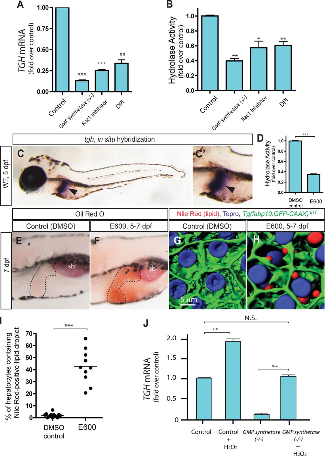
Fig. 5
Expression of the tgh gene is regulated by Rac1-mediated ROS. (A) qPCR analysis of tgh mRNA expression levels in wild-type control, GMP synthetases850 mutant, Rac1 inhibitor-treated, and DPI-treated larvae at 7 dpf. The averages of at least three independent experiments are shown. tgh mRNA expression level is significantly down-regulated in all tested conditions. (B) Hydrolysis activity toward p-nitrophenyl laurate was measured using lysates of control, GMP synthetases850 mutant, Rac1 inhibitor-treated, and DPI-treated larvae at 7 dpf. Measured activity was normalized to control and the average of three different experiments is shown. Hydrolysis activities are significantly decreased in all tested cases. (C) tgh expression in 5 dpf wild-type larvae. Lateral views, anterior to the left. The region around the liver is magnified and shown separately in C′. The expression of tgh is restricted to the liver at this stage. Arrowheads point to the liver. (D) The hydrolysis activity of the homogenate of 7 dpf larvae is measured in the absence or presence of E600. E600 significantly down-regulated hydrolysis activity. (E,F) Lateral views of DMSO- (C) or E600- (D) treated larvae stained for ORO at 7 dpf. Hydrolase inhibitor, E600, treated larvae (average 62.2%; SD 10.6; P < 0.001) developed hepatic steatosis. The black broken lines outline the liver. ORO staining experiments with E600-treated larvae were repeated six times with an average n = 14 larvae per experiment (total n = 84 larvae examined and total n = 52 larvae showed ORO signal in the liver). (G,H) Projected confocal images of lipid droplets in the liver stained by Nile Red. Tg (fabp10:GFP-CAAX)lri1 larvae treated with DMSO (G) or E600 (H) visualized for GFP expression and Nile Red (red) and To-pro-3 (blue) staining at 7 dpf. (I) Quantification of liver steatosis measured by the percentage of hepatocytes containing Nile Red-positive lipid droplets in DMSO- or E600-treated larvae at 7 dpf. E600-treated larvae developed hepatic steatosis. (J) qPCR analysis of tgh mRNA expression levels in wild-type, H2O2-treated wild-type, GMP synthetases850 mutant, and H2O2-treated GMP synthetases850 mutant larvae. tgh mRNA expression levels were restored to the level of wild-type control in H2O2-treated GMP synthetases850 mutant larvae. *P < 0.05, **P < 0.01, ***P < 0.001, n.s., not significant; error bars indicate SD.