
|
Fig. 1
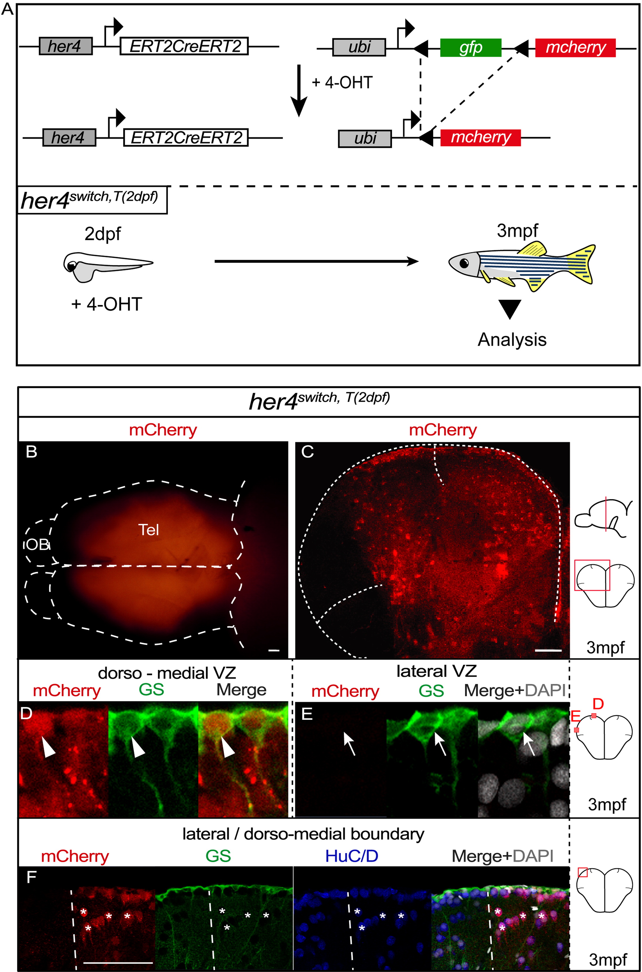
her4-Expressing Progenitors at 2 dpf Generate Adult NSCs of the Dorsomedial Pallium
(A) Genetic strategy used for the time-controlled fate mapping of her4-expressing progenitors: 4-hydroxy-tamoxifen (4-OHT) triggers ERT2CreERT2 activation in her4+ progenitors allowing GFP excision and permanent mcherry expression (top). Experimental design to map the adult fate of early her4+ progenitors: 4-OHT is applied at 2 dpf (her4switch,T(2 dpf)) and recombined animals are analyzed at 3mpf (bottom).
(B) Dorsal view (whole-mount, anterior left) of a her4switchT(2 dpf) adult telencephalon showing regionalized mCherry expression. Dotted lines delineate the telencephalon (Tel) and the olfactory bulb (OB).
(C) Cross-section of the telencephalon in a her4switch,T(2 dpf) adult, focusing on the pallium and stained as indicated. Dotted lines delineate pallial boundaries with the medial and lateral pallial sulci; one hemisphere is shown.
(D and E) High magnification of the dorsomedial (D) or lateral (E) NSCs. mCherry is expressed only in dorsomedial RGCs expressing GS (D, arrowheads) and not in lateral RGCs (E, arrows).
(F) Magnification of the boundary (dotted line) between the dorsomedial and lateral pallial domains, showing the segregation of mCherry+ and mCherry NSCs and neurons. Asterisks indicate some mCherry+ neurons.
See also Figure S1.
Reprinted from Developmental Cell, 30(2), Dirian, L., Galant, S., Coolen, M., Chen, W., Bedu, S., Houart, C., Bally-Cuif, L., Foucher, I., Spatial Regionalization and Heterochrony in the Formation of Adult Pallial Neural Stem Cells, 123-36, Copyright (2014) with permission from Elsevier. Full text @ Dev. Cell