
|
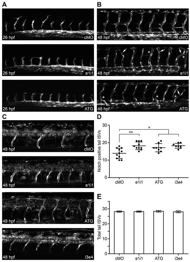
Fig. 6
papp-a2 knockdown causes angiogenesis defects and modulates vascular Notch signaling activity. (A,B) Representative z-stack projections of fluorescence microscopy images of control and papp-a2-knockdown Tg(fli1a:eGFP) zebrafish embryos at 26 hpf (A) and 48 hpf (B). Arrows, ISVs extending across somites; asterisks, abnormal ISV branching. (C) Representative z-stack projections of fluorescence microscopy images of control and papp-a2-knockdown embryos at 48 hpf in Notch activity reporter zebrafish embryos [Tg(Tp1bglob:eGFP)um13]. Knockdown was performed using 1.25ng ATG, 5.0ng e1i1, or 2ng i3e4 papp-a2-targeted morpholino per embryo. As a negative control, 5 ng cMO was injected. All pictures are lateral views, anterior to the left and dorsal to the top. (D) Number of Notch-signaling positive (the mean±s.d. is indicated) ISVs in the tails of 48 hpf control and papp-a2-knockdown embryos. *P<0.05, **P<0.01; n = 11 (cMO), 10 (e1i1), 8 (ATG), 8 (i3e4). (E) Total number of ISVs (the mean ± s.d. is indicated) in the tails of 48 hpf control and papp-a2 knockdown Tg(fli1a:eGFP) embryos. n = 6 per treatment.