
|
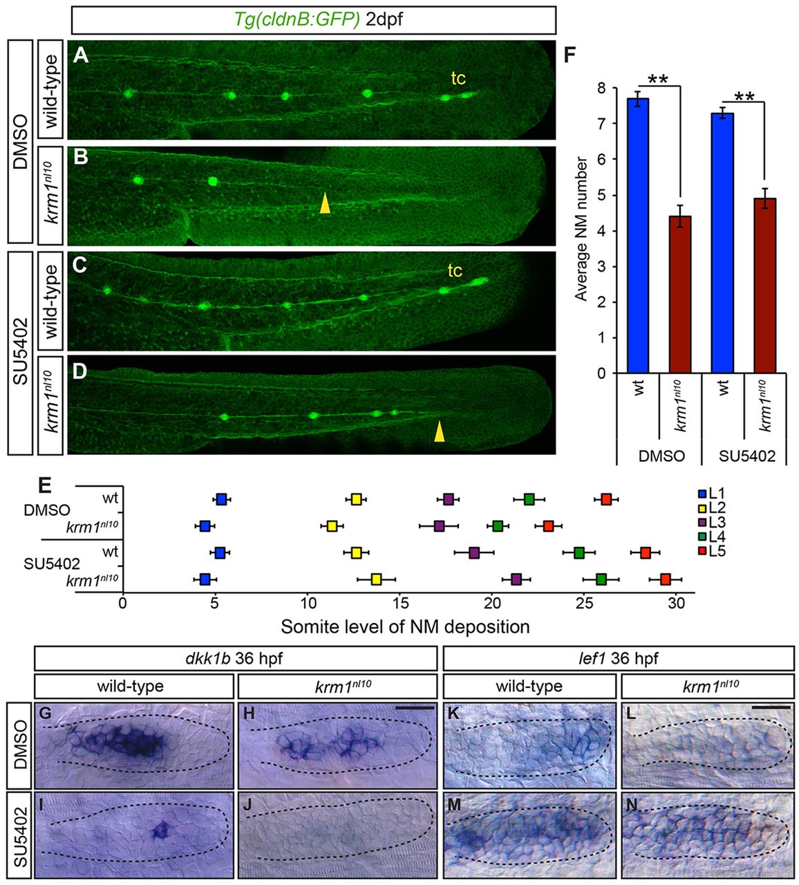
Fig. 6
Inhibition of Fgf signaling partially rescues NM spacing in krm1nl10 mutants. (A-D) Confocal projection of 2dpf Tg(cldnB:GFP) WT or krm1nl10 embryos, treated with either DMSO (controls) or SU5402. (A,E) DMSO-treated WT embryos show full pLLP extension and tc formation. (B,E) krm1nl10 mutants treated with DMSO show pLL truncation midway along the trunk (yellow arrowhead). (C,E) Suboptimal SU5402 treatment of WT embryos results in normal pLL extension and tc formation. (D,E) In krm1nl10 mutants, pLLP migration is partially rescued and pLL is extended to the end of the tail (yellow arrowhead), although tc NMs are rarely present (n=10 embryos per condition; P<0.001, two-way ANOVA with replication). (F) NM numbers are not significantly different between WT embryos treated with DMSO or SU5402 and between krm1nl10 mutants treated with DMSO or SU5402 (n=10 embryos/condition; **P<0.001, Student′s t-test). (G-N) Expression of dkk1b or lef1 in WT and krm1nl10 pLLPs treated with DMSO or SU5402 at 36 hpf. dkk1b is expressed in the mid-region of the pLLP of DMSO-treated WT (G) and mutant (H) embryos. In SU5402-treated embryos, dkk1b expression is reduced in WT pLLP (I) and absent in krm1nl10 pLLP (J). lef1 is expressed in the leading region of WT primordia (K) and absent in krm1nl10 mutant primordia treated with DMSO (L). SU5402 treatment results in increased lef1 expression in WT (M) and mutant (N) primordia. Scale bars: 20μm.