
|
Fig. 1
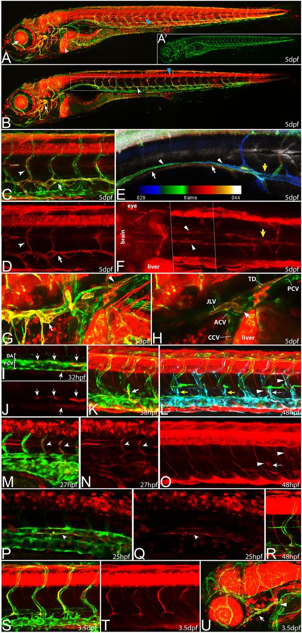
prox1a expression marks different endothelial compartments during vascular development. In all pictures (except for E), flt4:mCit expression is shown in green and prox1a:KalTA4,UAS:tagRFP expression is highlighted in red. (A) In full z-projections, prox1a expression at 5 dpf is apparent in various tissues, including liver (white arrowhead), lens (arrow) and myotome (blue arrowhead). (A2) Same z-projection displaying only the flt4:mCit expression in venous and lymphatic ECs. (B) Partial z-projection of the same embryo (comprising only optical sections without myotome signal) reveals additional expression in the spinal cord (blue arrowhead) and in the lymphatic vasculature of the head (arrow) and the trunk (white arrowhead). Images in A and B are composed of several overlapping z-projections because the embryo was too large to fit in a single field of view. (C,D) Higher magnification of trunk lymphatics at 5 dpf exhibiting combinatorial prox1a (red) and flt4 (green) reporter expression restricted to the TD (arrows) and ISLVs (arrowheads). (E) Depth color-coded z-projection (projecting each slice in a different color according to its position within the stack; see color bar) of boxed region in B (only prox1a channel) reveals the presence of two separate prox1a-positive vessels above the swim bladder (white arrows point to lymphatics on the right, arrowheads to lymphatics on the left body side), which merge near the sixth ISVs with the TD in the trunk region (yellow arrow). (F) Dorsal view of the prox1a-positive bilateral anterior TD (arrowheads), which connects to the trunk TD at the indicated location (yellow arrow). The image has been assembled from a set of partial z-projections (see dotted lines) owing to interference of other prox1a-positive structures. (G,H) Full (G) and partial (H) z-projections of the area (compare with box in A) where the anterior TD (arrowhead in G) and the facial lymphatic network (arrow in G) fuse and drain into the common cardinal vein (arrow in H) in a 5 dpf embryo. ACV, anterior cardinal vein; CCV, common cardinal vein; JLV, jugular lymphatic vessel; PCV, posterior cardinal vein; TD, thoracic duct. (I,J) At 32 hpf, prox1a-positive cells (red, arrows) are located within the PCV (green). Note that in this lateral view, prox1a-positive cells are located in both the dorsal and ventral aspect of the PCV. (K) A sprouting venous EC (arrow) at 38 hpf expressing both flt4 and prox1a transgenes. (L,O) At 48 hpf, only a proportion of PLs are prox1a:KalTA4-positive (white arrows) whereas the majority shows no signs of reporter expression (yellow arrows). In addition, venous ISVs positive for the prox1a reporter are evident (white arrowheads). (L) Overlay of a full z-projection of the flt4:mCit signal (green, to outline the complete vasculature) and identical partial z-projections of the prox1a (red, see also O) and flt4 reporter (blue, to highlight the part of the vasculature that is included in the partial z-projection of the prox1a signal). Note that prox1a-positive ECs will appear white in the overlay. (M,N) The prox1a reporter line also labels individual arterial sprouts (arrowheads) at 27 hpf. (P,Q) Single z-plane of the trunk region in a 25 hpf embryo showing DA cells positive for prox1a reporter expression (arrowheads). Note ventral domain expression of the DA. (R) Intersegmental vein showing both prox1a and flt4 reporter expression at 48 hpf. (S,T) At 3.5 dpf, all LECs display expression of the prox1a reporter but expression in other endothelial domains has disappeared. (U) Expression of prox1a in the head region at 3.5 dpf, highlighting the facial lymphatic network (arrow) including the drainage point with the common cardinal vein (arrowhead).