
|
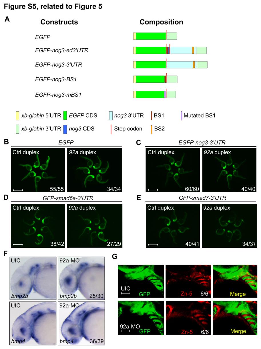
Fig. S5 Test of mir92a targets, related to Figure 5. (A) Schematic diagrams of reporter constructs. (B) The expression of the reporter GFP mRNA was unaffected by mir92a duplex. 60 pg of GFP mRNA was injected together with 1 nl of 20 M Ctrl duplex or mir92a duplex at the one-cell stage, and the injected embryos were observed for GFP fluorescence at 24 hpf. Scale bars, 1 mm. (C-E) mir92a had no effect on the expression of GFP-nog3-3′UTR (C), GFP-smad6a-3′UTR (D) and GFP-smad7-3′UTR (E) mRNAs. 60 pg of each mRNA species were co-injected with 1 nl of 20 M Ctrl duplex or mir92a duplex at the one-cell stage. The injected embryos were observed at 24 hpf. Scale bars, 1 mm. (F) bmp2b and bmp4 expression was detected by whole-mount in situ hybridization in uninjected control embryos (UIC) or mir92a morphants at 42 hpf. (G) Tg(fli1:GFP) transgenic embryos at 42 hpf were immunostained with anti-GFP and anti-Zn-5 antibodies. Scale bars, 50 µm.
Reprinted from Developmental Cell, 24(3), Ning, G., Liu, X., Dai, M., Meng, A., and Wang, Q., MicroRNA-92a upholds Bmp signaling by targeting noggin3 during pharyngeal cartilage formation, 283-295, Copyright (2013) with permission from Elsevier. Full text @ Dev. Cell