
|
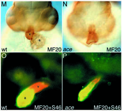
Fig. 1 Circulation, blood vessel formation and abnormal heart morphology in acerebellar mutants. (A,B) Overview of blood vessel system in a day-2 wild-type (A) and an ace (B) larva. (C,D) Confocal close-up of the blood vessels in the head in a wildtype (C) and an ace (D) larva on day 2, with the main vessels in the tectum, at the midhindbrain boundary and in the hindbrain being affected in ace mutants (arrows). The dashed circle in D marks the position of the eye. (E,F) The vessel at the mid-hindbrain boundary (E, arrowhead) is missing in ace mutant embryos (F, asterisk) at 24 hpf, detected by in situ hybridization with flk-1. aa, aortic arches; acv, anterior cardinal vein; h, heart; hbv, hindbrain vessel; mhbv, mid-hindbrain vessel; tv, tectal vessel; sv, segmental vessel. (G-J) Malformation of the heart in acerebellar larva (I) compared to wild type (G). G and I are lateral views of living larvae, H and J are schematic representation of the main structures in G and I. (K,L) Endocardium and myocardium are present and appear normal in ace mutants. (M,N) The heart is malformed and shortened in ace mutants, as shown by MF20 antibody staining (frontal view). MF20 antibody reacts with both cardiac chambers, whereas S46 specifically stains the atrium. (O,P) Double staining with MF20 and S46 facilitated measuring the chambers and the average length of ace hearts (83±16%, n=19) was found to be similar to the wild-type hearts (100±8%, n=10), whereas the ventricle was reduced in ace mutant embryos. In wild-type embryos, the ventricle contributes 39±4% (n=10) to the total heart length at 26 hpf, compared to only 24±7% (n=19) in ace mutants. Ventricle reduction becomes more pronounced, but is still variable at later stages: at 33 hpf, the ventricle is severely reduced or absent in 60% of all ace embryos (n=55), and less affected in the remaining embryos. (G-N) Day-2 larvae, (O,P) 26 hpf. a, atrium; bc, blood cells; e, endocard; h, heart; l, lens; m, myocard; p, pericard; v, ventricle; y, yolk.