
Image
|
Figure Caption
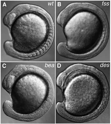
Fig. 3 Comparison of the three phenotypic groups of somite boundary mutants. All embryos are at the 11- to 13-somite stage. (A) Wild-type embryo. (B) fss mutant; only very few boundaries are present anteriorly. (C) bea mutant; in this individual the phenotype seems almost as severe as in fss; normally the anterior somites are more regular. (D) des mutant; the first eight somites look normal; no somites are formed beyond this point. The somite phenotype of aei and wit is similar to that of des.
Figure Data
Acknowledgments
This image is the copyrighted work of the attributed author or publisher, and
ZFIN has permission only to display this image to its users.
Additional permissions should be obtained from the applicable author or publisher of the image.
Full text @ Development