
|
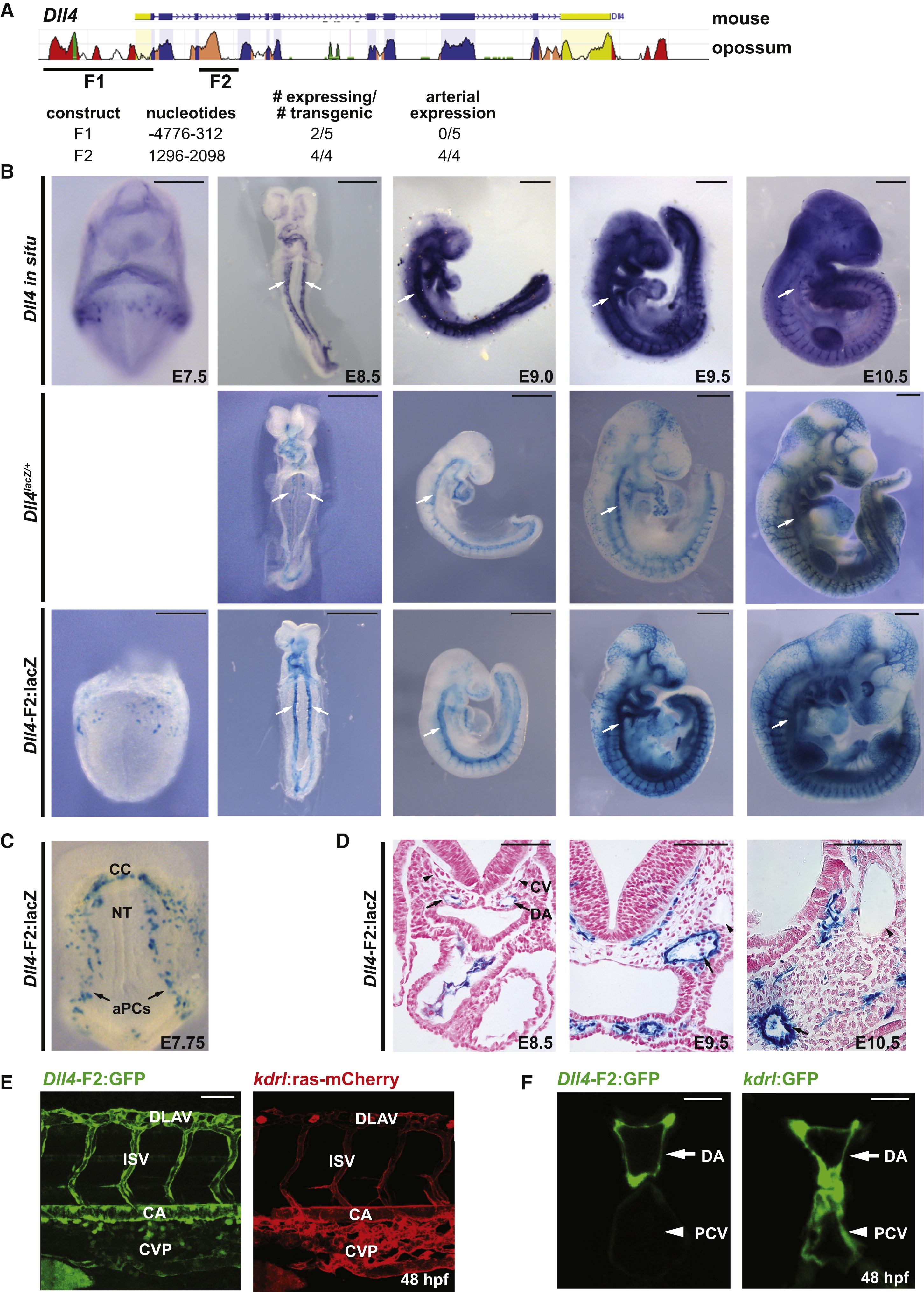
Fig. 1
Identification of an Intronic Enhancer of Dll4 that Drives Arterial-Specific Expression (A) Conservation between murine and opossum Dll4 with location of fragment 1 (F1) and F2 indicated. Transgenic analysis of F1-lacZ and F2-hsp68-lacZ (E9.5) is below. Further analysis of F1-lacZ is shown in Figures S1A and S1B. (B) In situ hybridization of endogenous Dll4 (top) and expression in Dll4lacZ/+ (middle) and a stable Dll4-F2-hps68-lacZ (F2) reporter line (bottom). Dorsal aorta (arrows). (C) F2 expression in early arterial precursors (aPCs) and in cardiac crescent (CC). NT, neural tube. (D) Transverse sections of F2 expression. DA, dorsal aorta (arrow); CV, cardinal vein (caret). (E) A stable Dll4-F2-E1b:GFP transgenic zebrafish line demonstrates arterial-specific expression. kdrl:ras-mCherry marks all blood vessels. CA, caudal artery; CVP, caudal vein plexus; ISV, intersomitic vessel; DLAV, dorsal longitudinal anastomotic vessel. (F) Cross-section of axial vasculature of F2:GFP zebrafish. PCV, posterior cardinal vein. Scale bars represent 500 μm (B), 100 μm (D), 50 μm (E), and 10 μm (F). See also Figure S1.
Reprinted from Developmental Cell, 26(1), Wythe, J.D., Dang, L.T., Devine, W.P., Boudreau, E., Artap, S.T., He, D., Schachterle, W., Stainier, D.Y., Oettgen, P., Black, B.L., Bruneau, B.G., and Fish, J.E., ETS Factors Regulate Vegf-Dependent Arterial Specification, 45-58, Copyright (2013) with permission from Elsevier. Full text @ Dev. Cell