
|
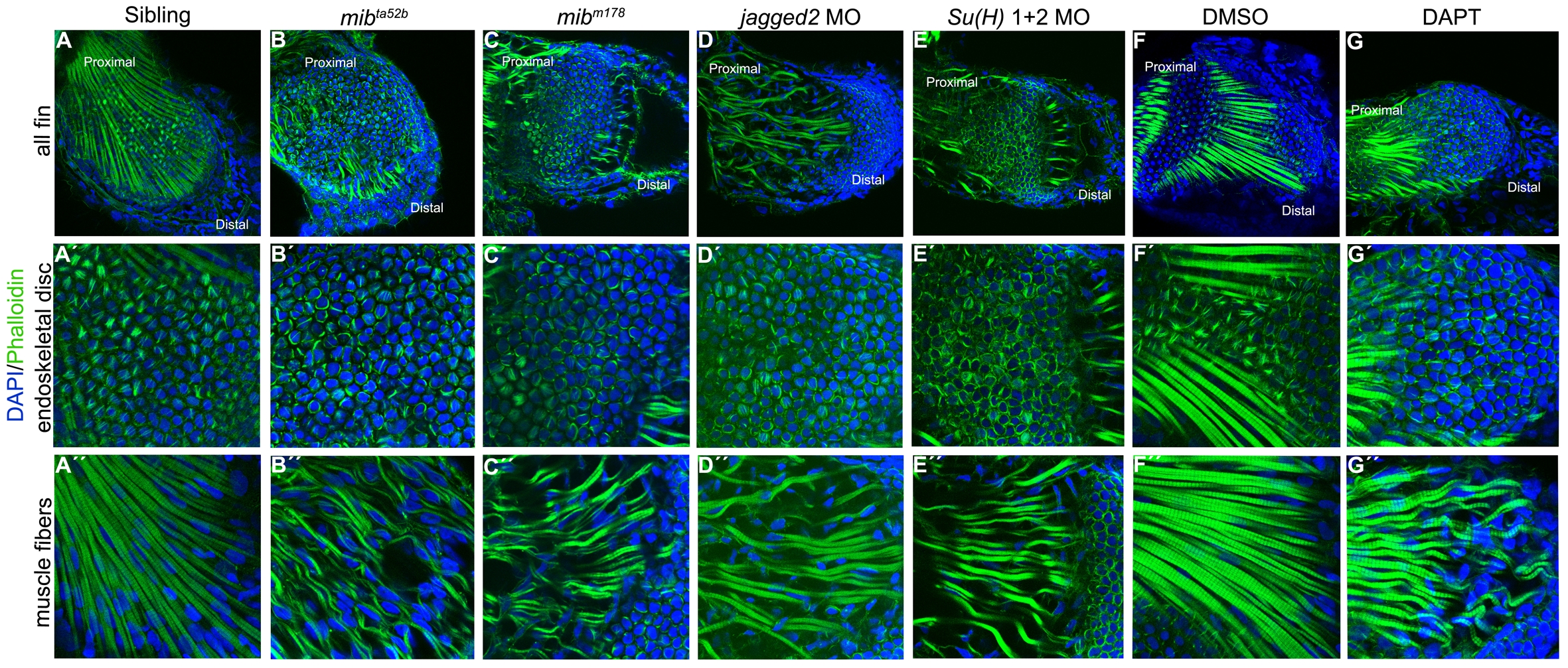
Fig. 4
Notch signalling is crucial for skeletal muscle integrity and stress fibres formation.
Immunostaining of 5 dpf pectoral fins (A–G′′) with DAPI and phalloidin to label nuclei (blue) and filamentous actin (green), respectively. Stress fibres in the endoskeletal disc cells and intact skeletal muscle fibres are formed in the pectoral fins of siblings (n = 20) (A–A′′) and in DMSO-treated embryos (n = 17) (F-F′′). In the pectoral fins were Notch signalling was disrupted like in mibta52b (n = 22) (B–B′′) and mibm178 (n = 10) (C–C′′) mutants, jagged2 (n = 11) (D–D′′) and Su(H)1+2 (n = 10) (E–E′′) morphants and in DAPT-treated embryos (n = 16) (G–G′′) the endoskeletal disc cells present high levels of actin at the periphery and the skeletal muscle fibres are wavy with gaps between them.