
|
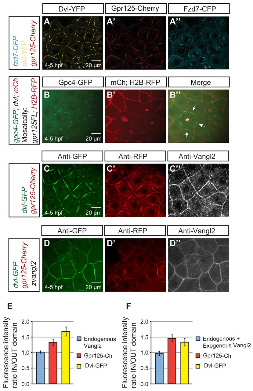
Fig. 7 Gpr125 promotes localization of select PCP components in Dvl-containing membrane subdomains in late blastulae (4-5 hpf). (A-A′) Live blastula co-injected with 110 pg fzd7-CFP, 150 pg dvl-YFP and 300 pg gpr125-Cherry RNA. (B-B′) Live blastula co-injected with 60 pg gpc4/kny-GFP, 150 pg dvl and 50 pg mCherry at the one-cell stage, and 4 pg H2B-RFP and 20 pg gpr125FL RNA in one blastomere at the 16- to <32-cell stage. The star in B′ marks an H2B-RFP-positive nucleus and arrows indicate membrane subdomains. (C-D′) Blastula injected with 300 pg gpr125-Cherry, 150 pg dvl-GFP (C-C′) and 50 pg zebrafish vangl2/tri RNA (D-D′) immunostained for GFP, RFP and Vangl2. Animal pole views in A-D. (E,F) Quantification of fluorescence intensity ratios inside/outside domain for Vangl2, Gpr125-Cherry and Dvl-GFP in embryos injected with 300 pg gpr125-Cherry, 150 pg dvl-GFP (E) and 50 pg zebrafish vangl2 RNA (F). Data are mean±s.e.m.