
Fig. 2
|
Fig. 2
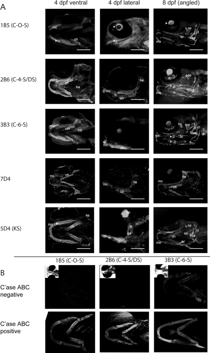
Various chondroitin sulfate moities are expressed in overlapping compartments during zebrafish skeletal development. A: Wholemount zebrafish heads labelled for various chondroitin and keratan sulfate epitopes with monoclonal antibodies 1B5, 2B6, 3B3, 7D4, and 5D4 (refer to Table 2, for antibody specificities). All images are confocal reconstructions presented with anterior facing to the left, and shown as ventral or lateral views. Scale bars = 100 μm in all panels. Triangular arrowheads in C-0-S and KS panels point to enrichment of GAG epitope in the jaw joint. Acute arrowheads in C-0-S panel point to expression in the lens. Acute arrowheads point to ossifying region of the ceratohyal. B: Chondroitinase ABC untreated controls show decreased/no immunoreactivity with the specific CS epitope antibodies. Treatment with chondroitinase ABC is required to generate the epitopes recognised by the 1B5 and 2B6 antibodies and, as such, chondroitinase untreated larvae show no immunoreactivity demonstrating that the antibodies are specific. The 3B3 antibody can also recognise an epitope present in native CS chains, so larvae untreated with chondroitinase ABC show changed rather than absent immunoreactivity with 3B3. mc, Meckel′s cartilage; ch, ceratohyal; ba, branchial arches; op, operculum; cl, cleithrum; 5ba, 5th branchial arch and teeth; nc, notochord; mx, maxilla; br, branchiosteal rays; hs, hyosymplectic; C′ase, chondroitinase.