
|
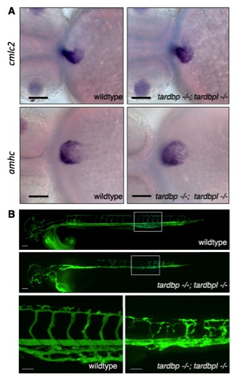
Fig. S4 In situ hybridization of cardiac markers and microangiography in tardbp-/-;tardbpl-/- mutants. (A) cmlc2 primarily stains the ventricle whereas amhc. primarily stains the atrial myocardium. These markers do not reveal any obvious differences in wild-type and tardbp-/-;tardbpl-/- embryos. (Scale bars, 100 μm.) Ventral view, anterior to the left, 2 dpf. (B) Green fluorescently labeled beads were microinjected into the lumen of the blood vessels (microangiography) to visualize the lumen of the vasculature. (Top) A wild-type embryo; (Middle) a tardbp-/-;tardbpl-/- mutant. (Scale bar, 100 μm.) (Bottom) Higher magnifications of the area in the white box of the upper panels. (Scale bars, 50 μm.) Lateral view, anterior to the left, 2 dpf.