
|
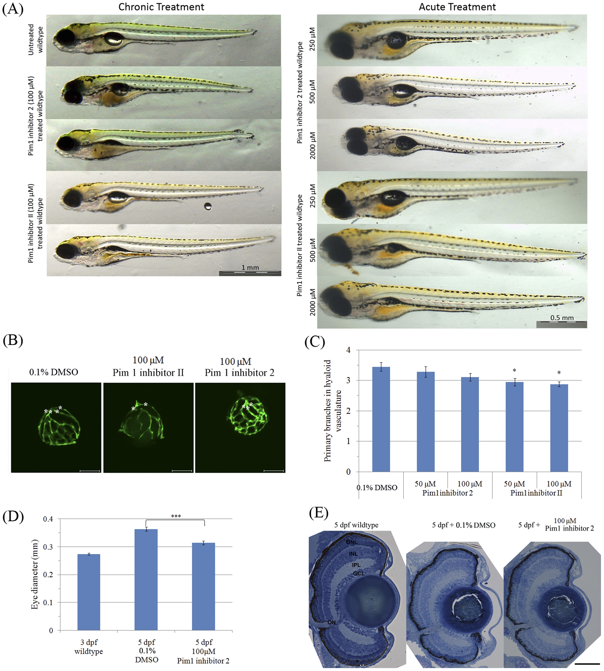
Fig. 8
Larvae treated with Pim1 inhibitors have a slightly reduced number of primary hyaloid vessels and eye size.
(A) Representative images of whole zebrafish larvae treated using Pim1 inhibitors. (B) and (C) zebrafish (n = 17 to 20) treated with Pim1 inhibitor 2 from 3–5 dpf have normal hyaloid vasculature morphology, while treatment with Pim1 inhibitor II from 3–5 dpf slightly reduces the number of primary hyaloid vessels. Primary hyaloid vessels are pointed using asterisks. P-value was calculated using one way ANOVA with Dunnett′s correction for multiple comparisons. *:ANOVA p<0.05. (D) Zebrafish larvae treated with Pim1 Inhibitor 2 from 3–5 dpf have a smaller eye. ***: Student′s t test p<0.001. (E) Retinal lamination appears normal in the larvae with drug-treated from 3–5 dpf. Scale bars are 1 mm (A) and 50 mm (E).