
|
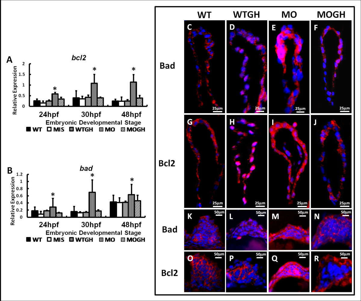
Fig. 4
Expression of apoptosis-related genes and proteins in tbx5 knockdown and growth hormone (GH)-treated zebrafish embryos at 30 h post-fertilization (hpf). Using a semiquantitative RT-PCR, the apoptotic genes, bcl2 (A) and bad (B), were significantly induced in MO group embryos and showed no significant differences among the WT, WTGH, and MOGH groups. (n = 3, 50 embryos/stage; relative expression = gene expression/β-actin expression). (C-R) Zebrafish embryos were stained by apoptosis-related antibodies, Bad and Bcl2 (red), and counterstained with DAPI (blue) for nucleus observation. In sagittal sections of the heart, Bad and Bcl2 were similarly expressed and significantly induced in tbx5-deficienct embryos (E, I), and showed no significant differences among the WT (C, G), WTGH (D, H), and MOGH (F, J) groups. Transverse sections showed that expression patterns of Bad and Bcl2 in pectoral fins were significantly induced in tbx5-deficient embryos (M, Q) and showed insignificant differences among the WT (K, O), WTGH (L, P), and MOGH (N, R) groups. (C-R) Embryo anteriors are to the left. WT, wild-type embryos; MO, tbx5 knockdown; MIS, mismatch tbx5-MO-treated embryos; WTGH, WT embryos treated with GH; MOGH, tbx5-MO- and GH-treated embryos. Data are presented as the mean ± S.D. *p < 0.05 vs. WT