
Image
|
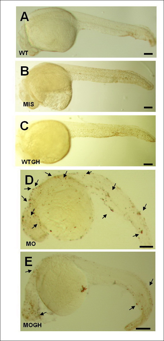
Figure Caption
Fig. 3 Growth hormone (GH)-treated tbx5 -knockdown zebrafish embryos show reduced apoptosis at 30 h post-fertilization (hpf). A TUNEL assay revealed no apoptotic spots in WT (A), MIS (B), and WTGH (C) embryos. (D) However, massive apoptotic spots were visible in MO embryos. (E) In the MOGH group, apoptotic sites were reduced. (A-E) Embryo anteriors are to the left. Scale bar = 0.1 cm. Black arrow, apoptotic site; WT, wild-type embryos; MO, tbx5 knockdown; MIS, mismatched tbx5-MO-treated embryos; WTGH, WT embryos treated with GH; MOGH, tbx5-MO- and GH-treated embryos.
Figure Data
Acknowledgments
This image is the copyrighted work of the attributed author or publisher, and
ZFIN has permission only to display this image to its users.
Additional permissions should be obtained from the applicable author or publisher of the image.
Full text @ J. Biomed. Sci.