
|
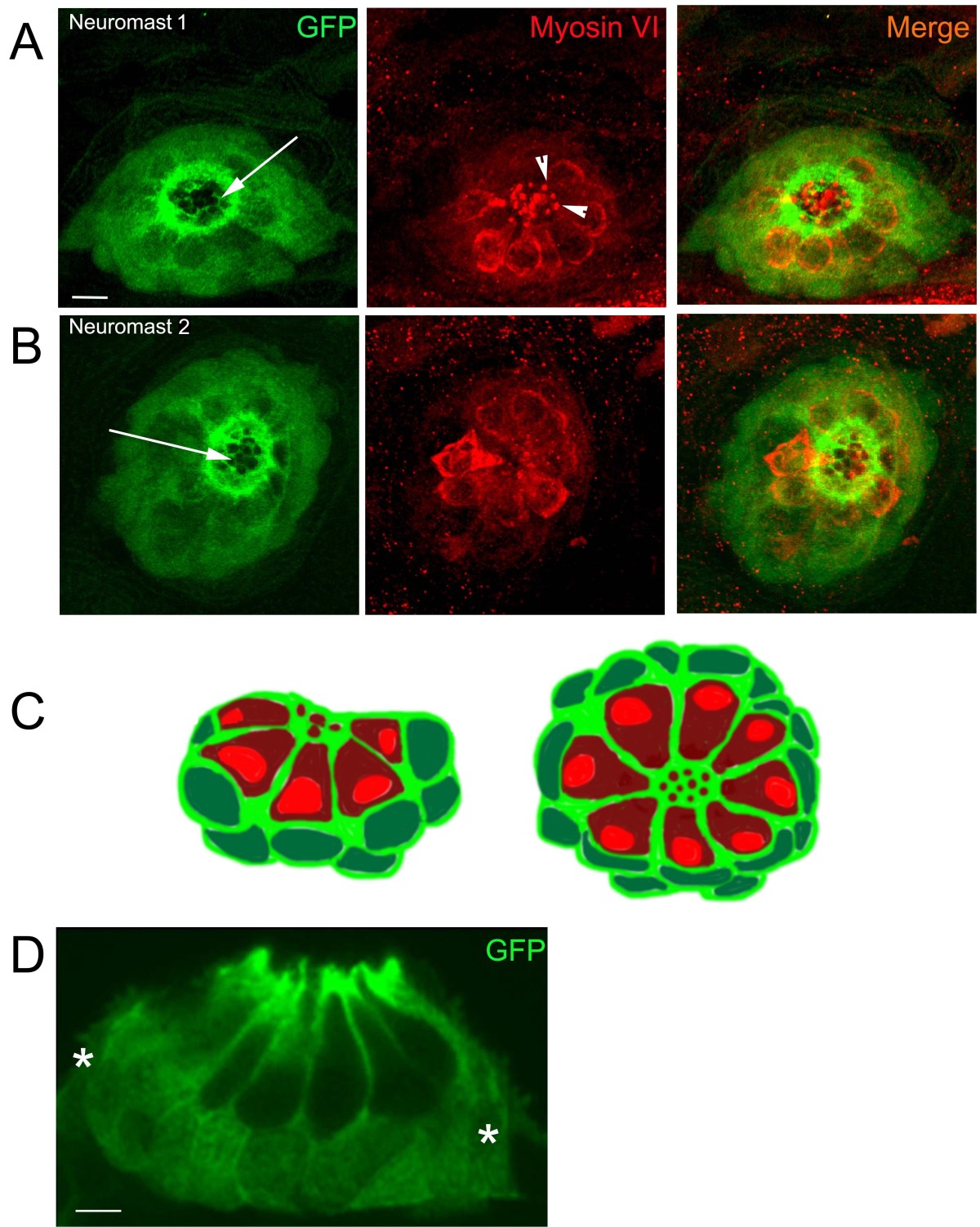
Fig. 2
Immuno-staining in neuromasts of 5 dpf Tg (tnks1bp1:EGFP) larvae. A. B. Confocal images of two different neuromasts (1 and 2) double-stained with anti-GFP (green, first and last columns) and anti-Myosin VI (red, second and last columns). The GFP positive cells (green) are the accessory cells (i.e. a combination of supporting cells and mantle cells). Around the apical pole of hair cells, accessory cells form a precisely organized honeycomb like annular structure (white arrows in A and B, first columns). Hair cells send their tightly packed hair bundles through the openings in the top, (white arrowheads in A, middle panel). C. Schematic cross-section (left image) and dorsal view (right image) of a neuromast illustrating the respective position of the accessory cells (cytoplasm light green and nucleus dark green) and the hair cells (cytoplasm dark red and nucleus light red). D. Cryosection of a neuromast, immunolabeled with an anti-GFP antibody. GFP expression is excluded from all hair cells (nucleus and cytoplasm) and found in all accessory cells, comprising the supporting and mantle cells (white stars). - 10 microns in A and B, 5 microns in D.