
|
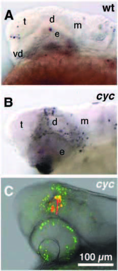
Fig. 6
Cells that fail to migrate in cyclops mutant embryos die. (A,B) bright field; (C) superimposed confocal and bright-field image; side views, anterior to left, dorsal to the top, 24h. (A) TUNEL labeling of wildtype embryo. Cell death is observed only in the surface ectoderm and the lens (out of focus). No cell death is apparent in forebrain or midbrain. Similar results were obtained in 19 other embryos. (B) TUNEL labeling reveals dying cells dorsal and posterior to the eye of cyclops mutant embryos (n=7). (C) Embryos (n=5) doubly labeled with lineage tracer (rhodamine dextran, red) and Acridine Orange (AO, green) showed that diencephalic precursors failed to move forward to separate the single retinal field. Lineage tracer injected cells remained posterior / dorsal to the eyes and died in dorsal diencephalic and pretectal regions (yellow, doubly labeled with AO and lineage tracer). A few hours later all rhodamine labeled cells were doubly labeled with AO (not shown). In wild-type embryos, lineage tracer labeled cells were unlabeled by AO (n=12). Prospective brain regions: t, telencephalon; vd, ventral diencephalon; d, dorsal/posterior diencephalon; e, eye; m, midbrain. Scale bar, 100 μm.