
Fig. 3
|
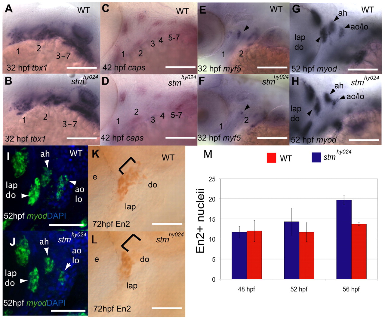
Fig. 3 stmhy024 mutants show myogenic defects specifically in opercular muscles. (A-H) In situ hybridisation with tbx1 (A,B) and capsulin (C,D) probes reveals that muscle specification is unaffected in the pharyngeal arch muscle precursors of stmhy024. However, both myf5 (E,F) and myod (G,H) expression are reduced in stmhy024 relative to wild type (WT). Arrowheads indicate muscle primordia. (I-L) Fluorescent in situ reveals that myod expression is reduced specifically in the precursors of the lo and ao at 52 hours post-fertilisation (hpf; I,J) but is unaffected in the adjacent ah. At later stages, there are fewer En2-expressing (En2+) muscle cells specifically in the do but not the lap of stmhy024 (K,L). Brackets indicate do muscle cell nuclei. (M) Quantification of En2+ muscle precursors in stmhy024 (blue) and WT (red) at 48, 52 and 56 hpf reveals that stmhy024 mutants have fewer muscle precursor cells than WT in the lap or do primordia from 56 hpf (mean ± s.e.m.). Pharyngeal arches 1-7 are shown by numbering in A-F. ah, adductor hyoideous; ao, adductor operculi; do, dilator operculi; e, eye; lap, levator arcus palatini; lo, levator operculi. Scale bars: 50 μm in A-H,K-L; 20 μm in I,J.