
|
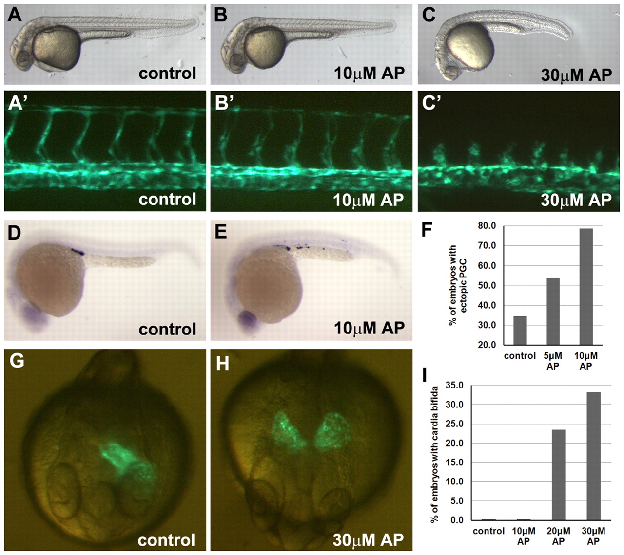
Fig. 8 Aplexone inhibits migration of germ cells, cardiomyocytes and arterial endothelial cells. (A,B,C) Overall phenotype of control and aplexone-treated embryos at 30 hpf. (A2,B′,C′) Vasculature in trunk. Aplexone (10 μM) does not affect the development of primary ISVs but 30 μM aplexone inhibits the growth of primary ISVs. (D,E) Germ cells in control (D) and aplexone-treated (E) embryos at 24 hpf were detected by in situ hybridization using the nanos probe. (F) The percentage of embryos with ectopic primordial germ cells in control (n=26), 5 μM (n=13) and 10 μM (n=14) aplexone-treated embryos. (G,H) Cardiomyocytes migrate to the midline and form a heart tube in Tg(myl7:GFP) embryos by 24 hpf (G), but fail to migrate to midline in embryos treated with 30 μM aplexone (H). (I) The percentage of embryos with cardia bifida in control (n=16) and 10 μM (n=17), 20 μM (n=17) and 30 μM (n=18) aplexone-treated embryos.