
|
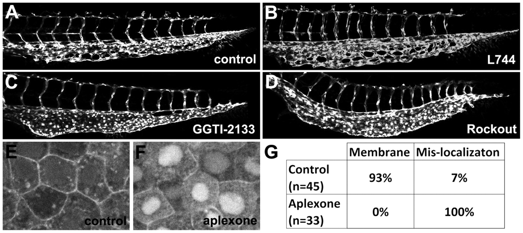
Fig. 6 Aplexone inhibits caudal vein angiogenesis by blocking geranylgeranylation. (A) Caudal vein of control Tg(kdrl:GFP) embryo at 48 hpf. (B-D) Caudal vein of L744,832-injected (B), GGTI-2133-injected (C) or 50 μM rockout-treated (D) Tg(kdrl:GFP) embryos analyzed at 48 hpf. (E,F) Representative confocal images of the localization of mCherry-rhoCAAX fusion proteins in control (E) and aplexone-treated (F) embryos at 80% epiboly. mCherry-rhoCAAX fusion proteins are predominantly localized to the plasma membrane in control embryos but are mislocalized to the nucleus and cytoplasm in aplexone-treated embryos. (G) Table quantifying the subcellular localization of mCherry-rhoCAAX in control or aplexone-treated embryos.