
|
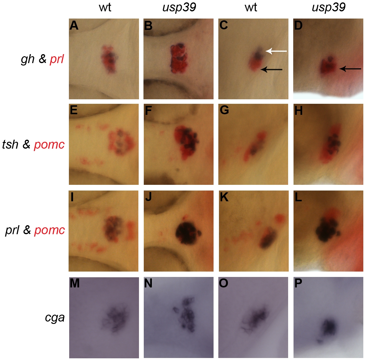
Fig. 3 usp39 mutation lead to expansion of all pituitary cell lineages at 48 hpf as indicated by pituitary hormone markers.
A–L: Whole-mount double in situ hybridization with probes indicated on the side. usp39 mutant embryos exhibit higher expression of all pituitary hormone markers compared to wild-type (wt) embryos. Spatial distribution of prl, tsh, pomc and cga are normal in the usp39 mutant. (A, B, E, F, I, J, M, and N) ventral view and (C, D, G, H, K, L, O, and P) lateral view, with anterior to the left. Columns 1 (A, E, I, and M) and 3 (C, G, K and O) show wt siblings; columns 2 (B, F, J, and N) and 4 (D, H, L, and P) show usp39 mutant embryos. A–D: gh (purple) and prl (red) transcripts. (C) The spatial distribution of gh is normally found in the proximal pars distalis (white arrow) and prl is found in the rostral pars distalis (black arrow). (D) Note the spatial distribution of gh in the usp39 mutant; gh is abnormally expressed in the rostral pars distalis (black arrow). E–H: tsh (purple) and pomc (red) transcripts. I–L: prl (purple) and pomc (red) transcripts. M–P: Whole-mount in situ hybridization with cga transcript.