
Fig. 3
|
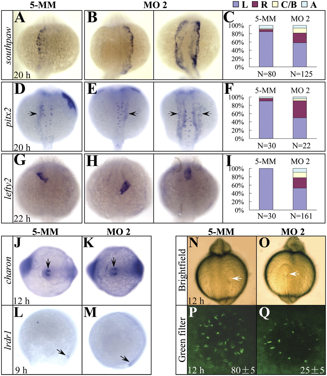
Fig. 3 The expression of asymmetric genes is randomized and cilia development in the Kupffer’s vesicle is disrupted in setdb2-knockdown embryos. (A–I) Whole-mount mRNA in situ hybridization and statistical analyses of the expression of asymmetric genes southpaw (A–C), pitx2 (D–F, arrows), and lefty2 (G–I) in the 5-MM control and setdb2 MO2-injected embryos at the indicated developmental stages. L, Left side; R, Right side; C/B, central or bilateral; A, absent. All embryos are dorsal views with head to the top. (J–M) WISH analyses of charon (J and K, arrows) and lrdr1 expression (L and M, arrows) in the 5-MM control and setdb2 MO2-injected embryos. (N–Q) Morphology of Kupffer’s vesicle (N and O, arrows) and confocol analyses of ciliary development (P and Q, green) as revealed by immunostaining with anti-acetylated tubulin antibody. The numbers in P and Q denote the mean ± SD. Embryos are ventral views in J, K, N, and O and lateral views in L and M.