
Fig. 4
|
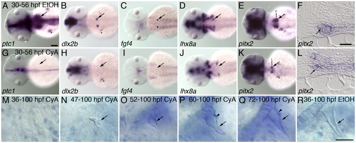
Fig. 4 Hedgehog requirements throughout tooth development. Inhibition of hedgehog signaling with cyclopamine (CyA) starting at 30 hpf prevents tooth morphogenesis, and treatments at subsequent stages reveal later hedgehog signaling requirements during tooth formation. (A-E) mRNA in situ hybridizations of pharyngeal tooth gene expression at 56 hpf for ptc1 (A), dlx2b (B), fgf4 (C), lhx8a (D), and pitx2 (E) in control embryos exposed to 0.5% EtOH from 30-56 hpf (dorsal views, anterior to the left, right side tooth germs indicated with an arrow, asterisks designate pectoral fin or girdle expression). (G-K) Embryos exposed to 50 μM CyA and 0.5% EtOH from 30-56 hpf show severely reduced or absent pharyngeal tooth expression of ptc1 (G), dlx2b (H), fgf4 (I), and lhx8a (L); but pitx2 (K) expression is maintained. (F, L) Transverse sections through the pharyngeal tooth forming region of 56 hpf embryos treated from 30 to 56 hpf in 0.5% EtOH with and without 50 μM CyA. Dental epithelial morphogenesis is highlighted by pitx2 mRNA expression in control embryos (F, arrow), but after CyA exposure the lower margin of the pharyngeal epithelium lacks the thickening or curved appearance characteristic of early tooth morphogenesis (L, arrow). (M-R) Right-side first pharyngeal tooth (arrows) in CyA or control treated, alcian blue stained and cleared 100 hpf larvae. No mature tooth formation was visible when CyA treatment was begun by 36 hpf (M). CyA exposure from 47 hpf allowed some limited mineralized morphogenesis, but it severely disrupted the shape of the entire tooth, causing it to appear unorganized (N). Treatment from 52 hpf resulted in teeth with a more regular appearance, but somewhat small and rounded (O). 60 hpf (P), and 72 hpf (Q), treatment resulted in teeth with only the later-developing shaft and base of the tooth having an abnormal rounded morphology (arrowhead) relative to controls (R). Scale bars: (A) 100 μm, (F) and (R) 25 μm.